The mass of the heart of an adult is approximately. Human heart weight
The structure of the heart of any organism has many characteristic nuances. In the process of phylogenesis, that is, the evolution of living organisms to more complex ones, the heart of birds, animals and humans acquires four chambers instead of two chambers in fish and three chambers in amphibians. Such a complex structure is best suited for the separation of arterial and venous blood flows. In addition, the anatomy of the human heart implies many tiny details, each of which performs its strictly defined functions.
Heart as an organ
So, the heart is nothing more than a hollow organ, consisting of specific muscle tissue, which performs the motor function. The heart is located in the chest behind the sternum, more on the left, and its longitudinal axis is directed anteriorly, to the left and down. In front, the heart borders on the lungs, almost completely covered by them, leaving only a small part directly adjacent to the chest from the inside. The boundaries of this part are otherwise called absolute cardiac dullness, and they can be determined by tapping the chest wall ().
In people with a normal constitution, the heart has a semi-horizontal position in the chest cavity, in people with an asthenic constitution (thin and tall) it is almost vertical, and in hypersthenics (dense, stocky, with large muscle mass) it is almost horizontal.

heart position
The back wall of the heart is adjacent to the esophagus and to large main vessels (to the thoracic aorta, to the inferior vena cava). The lower part of the heart is located on the diaphragm.

external structure of the heart
Age features
The human heart begins to form in the third week of the intrauterine period and continues throughout the entire period of gestation, passing through stages from a single-chamber cavity to a four-chamber heart.

development of the heart in utero
The formation of four chambers (two atria and two ventricles) occurs already in the first two months of pregnancy. The smallest structures are fully formed by childbirth. It is in the first two months that the heart of the embryo is most vulnerable to the negative influence of certain factors on the expectant mother.
The heart of the fetus is involved in the blood flow through his body, but differs in the circles of blood circulation - the fetus does not yet have its own breathing with the lungs, but it “breathes” through the placental blood. There are some openings in the fetal heart that allow the pulmonary blood flow to be "switched off" from circulation prior to birth. During childbirth, accompanied by the first cry of the newborn, and, therefore, at the time of increased intrathoracic pressure and pressure in the heart of the child, these openings are closed. But this does not always happen, and they may remain in a child, for example, (not to be confused with such a defect as an atrial septal defect). An open window is not a heart defect, and subsequently, as the child grows, it overgrows.

hemodynamics in the heart before and after birth
The heart of a newborn child has a rounded shape, and its dimensions are 3-4 cm in length and 3-3.5 cm in width. In the first year of a child's life, the heart increases significantly in size, and more in length than in width. The mass of the heart of a newborn child is about 25-30 grams.
As the baby grows and develops, the heart also grows, sometimes significantly outpacing the development of the body itself according to age. By the age of 15, the mass of the heart increases by almost ten times, and its volume increases by more than five times. The heart grows most intensively up to five years, and then during puberty.
In an adult, the heart is about 11-14 cm long and 8-10 cm wide. Many rightly believe that the size of the heart of each person corresponds to the size of his clenched fist. The mass of the heart in women is about 200 grams, and in men - about 300-350 grams.
After 25 years, changes begin in the connective tissue of the heart, which forms the heart valves. Their elasticity is no longer the same as in childhood and adolescence, and the edges may become uneven. As a person grows up, and then aging, changes occur in all structures of the heart, as well as in the vessels that feed it (in the coronary arteries). These changes can lead to the development of numerous cardiac diseases.
Anatomical and functional features of the heart
Anatomically, the heart is an organ divided by partitions and valves into four chambers. The “upper” two are called the atria (atrium), and the “lower” two are called the ventricles (ventriculum). Between the right and left atria is the interatrial septum, and between the ventricles is the interventricular septum. Normally, these partitions do not have holes in them. If there are holes, this leads to mixing of arterial and venous blood, and, accordingly, to hypoxia of many organs and tissues. Such holes are called septal defects and refer to.

basic structure of the chambers of the heart
The boundaries between the upper and lower chambers are atrioventricular openings - the left, covered by the leaflets of the mitral valve, and the right, covered by the leaflets of the tricuspid valve. The integrity of the septa and the proper functioning of the valvular leaflets prevent mixing of blood flows in the heart, and promote a clear unidirectional flow of blood.
The atria and ventricles are different - the atria are smaller than the ventricles and have thinner walls. So, the wall of the atria is about only three millimeters, the wall of the right ventricle is about 0.5 cm, and the left one is about 1.5 cm.
The atria have small protrusions - ears. They have a slight suction function for better pumping of blood into the atrial cavity. The mouth of the vena cava flows into the right atrium near its ear, and the pulmonary veins in the amount of four (rarely five) flow into the left atrium. From the ventricles depart the pulmonary artery (more often called the pulmonary trunk) on the right and the aortic bulb on the left.

structure of the heart and its vessels
From the inside, the upper and lower chambers of the heart also differ and have their own characteristics. The surface of the atria is smoother than that of the ventricles. From the valve ring between the atrium and the ventricle, thin connective tissue valves originate - bicuspid (mitral) on the left and tricuspid (tricuspid) on the right. The other edge of the leaflet faces the inside of the ventricles. But in order for them not to hang freely, they are, as it were, supported by thin tendon threads called chords. They are like springs, stretch when the valve flaps close and contract when the flaps open. Chords originate from the papillary muscles from the wall of the ventricles - three in the right and two in the left ventricle. That is why the ventricular cavity has an uneven and bumpy inner surface.
The functions of the atria and ventricles also differ. Due to the fact that the atria need to push blood into the ventricles, and not into larger and longer vessels, they have less resistance to muscle tissue to overcome, so the atria are smaller in size and their walls are thinner than those of the ventricles. The ventricles push blood into the aorta (left) and into the pulmonary artery (right). Conventionally, the heart is divided into right and left halves. The right half serves for the flow of exclusively venous blood, and the left half for arterial blood. Schematically, "right heart" is indicated in blue, and "left heart" in red. Normally, these streams never mix.

hemodynamics in the heart
One cardiac cycle lasts about 1 second and is carried out as follows. At the moment of filling with blood, the walls of the atria relax - atrial diastole occurs. The valves of the hollow veins and pulmonary veins are open. The tricuspid and mitral valves are closed. Then the atrial walls tighten and push blood into the ventricles, the tricuspid and mitral valves open. At this point, there is systole (contraction) of the atria and diastole (relaxation) of the ventricles. After the ventricles have taken in blood, the tricuspid and mitral valves close, and the aortic and pulmonary valves open. Then the ventricles contract (ventricular systole), and the atria fill with blood again. There comes a general diastole of the heart.

cardiac cycle
The main function of the heart is reduced to pumping, that is, to pushing a certain blood volume into the aorta with such pressure and speed that the blood is delivered to the most distant organs and to the smallest cells of the body. Moreover, arterial blood with a high content of oxygen and nutrients is pushed into the aorta, which enters the left half of the heart from the vessels of the lungs (flows to the heart through the pulmonary veins).
Venous blood, with a low content of oxygen and other substances, is collected from all cells and organs from the vena cava system, and flows into the right half of the heart from the superior and inferior vena cava. Further, venous blood is pushed out of the right ventricle into the pulmonary artery, and then into the pulmonary vessels in order to carry out gas exchange in the alveoli of the lungs and to enrich it with oxygen. In the lungs, arterial blood collects in the pulmonary venules and veins, and again flows into the left half of the heart (into the left atrium). And so regularly the heart pumps blood around the body at a frequency of 60-80 beats per minute. These processes are denoted by the concept "Circulation of blood". There are two of them - small and large:
- small circle includes the flow of venous blood from the right atrium through the tricuspid valve into the right ventricle - then into the pulmonary artery - then into the arteries of the lungs - oxygenation of blood in the pulmonary alveoli - the flow of arterial blood into the smallest veins of the lungs - into the pulmonary veins - into the left atrium.
- big circle includes the flow of arterial blood from the left atrium through the mitral valve to the left ventricle - through the aorta into the arterial bed of all organs - after gas exchange in tissues and organs, the blood becomes venous (with a high content of carbon dioxide instead of oxygen) - further into the venous bed of organs - into the system of hollow veins - in the right atrium.

circles of blood circulation
Video: heart anatomy and cardiac cycle briefly
Morphological features of the heart
If you look at sections of the heart under a microscope, you can see a special type of musculature that is no longer found in any organ. This is a type of striated muscle, but with significant histological differences from ordinary skeletal muscles and from the muscles lining the internal organs. The main function of the heart muscle, or myocardium, is to provide the most important ability of the heart, which forms the basis of the vital activity of the whole organism as a whole. Is it the ability to shrink, or contractility.
In order for the fibers of the heart muscle to contract synchronously, electrical signals must be supplied to them, which excite the fibers. This is another capacity of the heart – .
Conductivity and contractility are possible due to the fact that the heart autonomously generates electricity in itself. Function Data (automatism and excitability) are provided with special fibers that are an integral part of the conductive system. The latter is represented by electrically active cells of the sinus node, the atrioventricular node, the His bundle (with two legs - right and left), as well as Purkinje fibers. In the case when a patient's myocardial damage affects these fibers, they develop, otherwise called.

cardiac cycle
Normally, an electrical impulse originates in the cells of the sinus node, which is located in the zone of the right atrial appendage. In a short period of time (about half a millisecond), the impulse propagates through the atrial myocardium, and then enters the cells of the atrioventricular junction. Usually, signals are transmitted to the AV node through three main tracts - the Wenckenbach, Thorel and Bachmann bundles. In the cells of the AV node, the time of impulse transmission is extended to 20-80 milliseconds, and then the impulses enter through the right and left legs (as well as the anterior and posterior branches of the left leg) of the His bundle to the Purkinje fibers, and eventually to the working myocardium. The frequency of impulse transmission along all pathways is equal to the heart rate and is 55-80 impulses per minute.
So, the myocardium, or cardiac muscle, is the middle membrane in the wall of the heart. The inner and outer shells are connective tissue, and are called the endocardium and epicardium. The last layer is part of the pericardial sac, or cardiac “shirt”. Between the inner sheet of the pericardium and the epicardium, a cavity is formed, filled with a very small amount of fluid, to ensure better sliding of the sheets of the pericardium at the moments of heart contractions. Normally, the volume of fluid is up to 50 ml, an excess of this volume may indicate pericarditis.

structure of the heart wall and membrane
Blood supply and innervation of the heart
Despite the fact that the heart is a pump to provide the whole body with oxygen and nutrients, it itself also needs arterial blood. In this regard, the entire wall of the heart has a well-developed arterial network, which is represented by a branching of the coronary (coronary) arteries. The mouths of the right and left coronary arteries depart from the aortic root and are divided into branches penetrating the thickness of the heart wall. If these important arteries become clogged with blood clots and atherosclerotic plaques, the patient will develop and the organ will no longer be able to perform its functions in full.

the location of the coronary arteries that supply blood to the heart muscle (myocardium)
The frequency and strength with which the heart beats is influenced by nerve fibers extending from the most important nerve conductors - the vagus nerve and the sympathetic trunk. The first fibers have the ability to slow down the frequency of the rhythm, the latter - to increase the frequency and strength of the heartbeat, that is, they act like adrenaline.
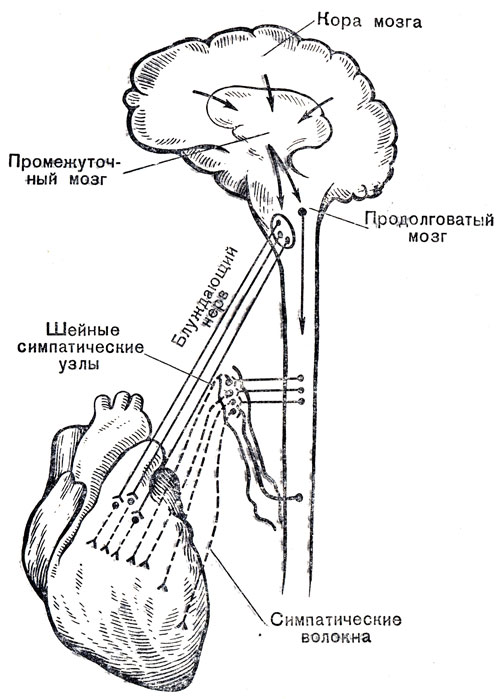
innervation of the heart
In conclusion, it should be noted that the anatomy of the heart may have some deviations in individual patients, therefore, only a doctor is able to determine the norm or pathology in a person after conducting an examination that can most informatively visualize the cardiovascular system.
Video: lecture on the anatomy of the heart
The dimensions of the fetal heart can be examined starting from the 20th week of pregnancy. After birth, it has the shape of a ball, and then by adolescence acquires an anatomical structure, as in adults. In diseases of the nervous, endocrine and digestive systems, body weight and, accordingly, the size of the heart may decrease. Microcardia is also a congenital pathology.
📌 Read this article
What is the size of the heart, valves and chambers in a person is normal
You can study the structure of the heart using ultrasound. Its size is determined by age, physique. With malformations, there are violations - a deviation up or down. The data obtained during the instrumental study are needed to make a diagnosis and choose the tactics of treating the patient.
In the fetus
You can hear the work of the heart at week 8, but it is possible to detect the atria and ventricles in it only closer to the twentieth. It is usually recommended to have an ultrasound to measure the fetal heart by 24 weeks of gestation. The organ itself is easily detected in the chest cavity, but the study of its structure and volume can be difficult, especially with high fetal mobility or a decrease in the amount of amniotic fluid.
With the normal development of the heart, its characteristics are:
- location in the left half of the chest;
- the shape of a hollow ball;
- detection of four chambers, valves between them, septum, aortic arch.

 Ultrasound of the fetal heart (color doppler)
 Since the heart of the fetus resembles a ball, the main measurements are taken in any of the directions. The diameter at week 24 is 2.5 cm, and by birth it increases to 4.5. All these indicators are average, they depend on the weight of the child. The valves are visualized, their range of motion can be assessed.
The thickness of the heart muscle is also considered an important parameter - during contraction it is 4 mm, and in the relaxation phase - 2.9 mm. An essential feature of the fetal heart is the equal size of the ventricles.
In children and adolescents
As the child grows, the heart gradually acquires the features of an adult. Finally, this process is completed only by 11-14 years. In one-year-old children, the weight of the organ increases by 2 times compared with the parameters after childbirth, and by the age of three it is 3 times higher. By the age of 5-6, growth slows down a little, and in adolescents it accelerates again. In a 17-year-old young man or girl, the size of the myocardium is 10 times larger than in newborns.
Initially, the left ventricle increases, since it bears the main burden of pumping blood. Its size in a four-month-old baby is 2 times larger than in the right one. The thickness of the myocardium from 5 mm reaches 12 (left) and 6 mm (right). The relative volume of the heart (compared to the chest) is greater in children than in adults.
The heart of boys up to 10 years is larger than that of girls, then increased development by the age of 16 allows girls to get ahead of boys, then growth slows down again.
In one year, the heart has the following average parameters (diameter in cm):
- ventricular diameter - left 3.2 and right 1.4 (at maximum filling);
- atrium - left 2.4 and right 1.1;
- partition - 0.5.
For adolescents 15 years old, the left half of the heart normally has the following dimensions:
- end diastolic diameter of the ventricle 4.3 cm, systolic - 3.5 cm;
- left atrium - 3 cm;
- aortic diameter - from 2 to 3 cm.
The right ventricle at the same time reaches only 1.8 cm.
In an adult
The length of the heart varies from 9 to 16 cm. Most often it is close to 12.5 cm. The base of the heart is about 10.5 cm wide, and the size from the front to the back wall is 6 - 7.5. Heart chamber parameters (cm):
- diameters - left ventricle about 4.6, right 1.95, left atrium 2.9 - 3.1, right 1.9 - 2.5;
- valve circles - aorta 0.8 - 0.85 and pulmonary artery 0.57 - 0.98, and atrioventricular averages are about 1 cm;
- the thickness of the wall of the ventricles - right up to 0.5, and left up to 1.5.
Expert opinion
Alena Ariko
Expert in cardiology
At the same time, in well-trained athletes, the heart may be larger than normal, and in short, thin women, its size is lower. If there are no functional impairments, then such deviations can be neglected. Diagnosis is not made on the basis of echocardiographic or radiographic measurements alone. Clinical symptoms, laboratory data, are taken into account.
If they put small sizes - is it bad?
A decrease in the anatomical parameters of the chambers of the heart or the entire organ can be in the following conditions:
- Malformations- a severe form of cardiac hypoplasia. The left or right ventricle may be smaller. Usually associated with other congenital anomalies. If the entire left half of the heart is underdeveloped, it rapidly progresses with a fatal outcome.
- With a decrease in the right half, breathing is disturbed, cyanosis of the skin is noted, the main load falls on the left ventricle, with its weakening, stagnant processes in the internal organs increase.
- Exhaustion, protein-energy starvation- occurs with prolonged and severe infections, disruption of the digestive system, endocrine system, brain damage, malignant neoplasms. Against the background of weight loss, the size of the heart decreases.
- - less than the norm can only be the capacity of the chambers of the heart due to the excessive growth of the inner shell. It occurs with a genetic predisposition, diabetes, sarcoidosis, tumor processes, radiation therapy. Due to low cardiac output, circulatory failure occurs.
- - compaction of the sheets of the pericardial sac during inflammation compresses the heart from the outside. After the deposition of calcium salts, a strong shell is formed - a shell-like heart. This condition disrupts the filling of the ventricles, leads to exhaustion, disruption of the liver, edema, atrophy of muscle fibers, and arrhythmias.
For children, insufficient heart size compared to age norms is one of the signs of a lag in growth and development. This happens if:
- both parents or one of them is short;
- There is ;
- the child is diagnosed with a disease of the respiratory system;
- impaired function of the liver, digestive organs;
- there is a genetic anomaly;
- the pituitary gland, hypothalamus, thyroid gland are affected;
- has diabetes.
In elderly patients, as well as in old age, involutional processes are observed, the outcome of which may be atrophy of the heart muscle. This condition is an extreme manifestation of myocardial malnutrition (), which increases with aging of the body. Myocardial dystrophy can also accompany:
The size of the heart is determined by age, body type and the presence of diseases. A decrease occurs with congenital malformations of the structure, protein starvation, lagging behind children in growth and development. Restrictive cardiomyopathy or constrictive pericarditis can also cause such a change. In elderly patients, a possible cause is myocardial dystrophy or atrophy of the heart with sudden weight loss, metabolic disorders.
Useful video
Watch the video on how the human heart works:
Read also
An enlarged heart is not always indicative of pathology. Nevertheless, a change in size may indicate the presence of a dangerous syndrome, the causes of which are myocardial deformation. The symptoms are washed away, the diagnosis includes x-rays, fluorography. Treatment of cardiomegaly is long, the consequences may require a heart transplant.

in women 1:180. And many more interesting facts about the human body can be found
How much does an adult human heart weigh? What is the size of a human heart?
What is the weight of the human heart?
How much does a human heart weigh?
A man's heart is roughly equal to the size of his fist. The weight of the heart of an adult is 220-260 g, and the ratio of the weight of the heart
(to the total body weight) is equal to men 1:170,
in women - 1:180. And many more interesting facts about the human body can be found
Infant heart weight at birth. After 8 months, it becomes twice as large. In an adult man, the heart weighs 300 grams, in a woman a few less. Well, about the fact that the size of a person's heart is equal to the size of his fist, I think everyone knows.
The average weight of the heart of an adult is g. The length of the heart is cm, width cm, height (thickness) cm. The thickness of the ventricles: left - 0.8 - 1 cm, right - about 0.5 cm.
Man → What is the largest human organ and what is the average weight of the human brain?
The names of the fingers of the French: pus, index, major, anulare, oryculaire.
Each human finger bends about 25 million times during its lifetime.
The size of a human heart is approximately equal to the size of his fist.
The weight of an adult human heart is
The composition of the human body includes only 4 minerals: apatite, aragonite, calcite and cristobalite.
The human brain generates more electrical impulses in a day than all the phones in the world combined.
The average human brain weighs 1.4 kilograms, while the size in this case really does not matter: Einstein's brain weighed 0.2 kg less than the norm, so if a friend's hat is small for you, don't flatter yourself - it's not a fact that you are smarter than him .
Despite the small volume and the fact that the brain is 90% water, it consumes 25% of all oxygen and sugar needed by the body.
The phenomenon in which a person loses the ability to see from strong light is called “snow blindness”.
The total weight of bacteria living in the human body is 2 kilograms.
Chemical reactions take place in the human brain in one second.
Children are born without kneecaps. They appear only at the age of 2-6 years.
From the moment of birth, there are already 14 billion cells in the human brain, and this number does not increase until death. On the contrary, after 25 years it is reduced by 100 thousand per day. About 70 cells die per minute you spend reading a page. After the age of 40, brain degradation accelerates sharply, and after 50, neurons (nerve cells) dry out and the volume of the brain shrinks.
In psychiatry, the syndrome, accompanied by depersonalization, a violation of the perception of time and space, one's own body and the environment, is officially (!) Called "Alice in Wonderland."
How to fix it? Either reduce oxygen consumption (lose weight, which is not acceptable), or increase the volume of the heart and blood distilled at a time. This, in fact, is the meaning of training the heart - to increase its internal volume.
The larger the volume of the heart, the more nutrients the heart receives at a time.
The larger the volume of the heart, the less often it can contract.
The less often the heart contracts (works), the less it wears out.
L and D - cardiac hypertrophy.
Note that I said an increase in the volume of the heart, not an increase in the size of the heart. These are very important things. Because the first is very useful, and the second, on the contrary, is very harmful! The fact is that cardiac hypertrophy can be good and bad. When the increase in volume occurs due to the stretching of the walls of the heart muscle (L-hypertrophy) - this is very good! This allows us to pump more blood at a time - which is what we need. But when the heart grows due to the thickening of the walls of the heart muscle (D - hypertrophy) - this is very bad. This is the so-called myocardial hypertrophy due to a diastolic defect. In general, such an unpleasant thing as a heart attack is the consequences of just such changes in the heart.
Fine. How to achieve good hypertrophy and avoid bad? Everything is very simple. No need to work in a pulse close to the maximum (beats)! You need to work long and often at an average heart rate () beats per minute. For the majority, pulse beats per minute are most often ideal. An ordinary healthy person at rest has a pulse of 70 beats per minute. When such a person begins to do some kind of cyclic long-term work (training with iron, running or walking fast), his pulse begins to increase in order to supply all the organs of the body with an increased amount of oxygen due to the load. Here his pulse reached 130 beats per minute. A person in this situation can stabilize the load and continue to work without increasing the intensity. If he continues this training for an hour, then the "flexibility" of his heart will begin to improve. Muscles will distill a huge amount of blood through the heart and it will begin to gradually stretch. If you train like this often (from 3 times a week for 60 minutes), then over time the heart will stretch and its volume will increase significantly. Accordingly, the volume of blood pumped per pulse will increase, endurance will increase, and the number of pulse beats at rest will decrease.
Heart medications work well with supplements such as St. John's wort, garlic, and ginkgo biloba.
Every day, the heart produces enough energy to drive a car 32 km. In a lifetime, this is the equivalent of going to the moon and back.
Cocaine affects the electrical activity of the heart and causes spasm of the arteries, which can lead to a heart attack even in healthy people on the planet.
It has always been believed that men are more prone to heart problems. But that's not the case at all. Here is a statistic to prove the fact: every year in the United States, 10 times more women die of heart disease than men.
A kitchen faucet must be kept fully open for 45 years to pour out an amount of water equal to the volume of blood pumped by the heart in an average human life.
Computed tomography of mummies showed that heart disease was surprisingly common in Egypt. Probably, in those days there were other factors that lead to heart disease, not including smoking, fast food and excessive television.
Single people have twice the risk of death from sudden cardiac arrest and myocardial infarction than those who live with relatives or at least have a roommate.
The first heart transplant was performed in 1967 by Dr. Christian Barnard from South Africa. The human heart was transplanted into the body of Louis Vashansky. Although the patient lived only 18 days after the operation, this case is considered the first successful heart transplant operation.
Galen of Pergamon, a prominent Roman gladiator surgeon, proved that the arteries were filled with blood and not air, as Hippocrates had previously assumed. However, he also believed that the heart acts as a low-temperature furnace that holds the heat of the blood and pumps it from one part of itself to another through tiny holes.
In most people, erythrocytes consist of normal hemoglobin A, but in a part of the population they are made of hemoglobin S. Such erythrocytes are crescent-shaped instead of round, which makes them less able to tolerate oxygen, have reduced resistance, and many other disadvantages. At the same time, there are quite a few carriers of sickle cell anemia, especially in regions of tropical and subtropical climates, where malaria is common.
It turns out that hemoglobin S significantly reduces the likelihood of contracting malaria, which, within large populations, prevents the degeneration of the mutant gene associated with it.
The amount of blood in the body, although it is a variable value for different people (about 7 percent of body weight), but does not exceed 7-10 liters.
Frozen red blood cells can be stored for up to 10 years.
The first blood banks appeared during the Second World War. Their founder is considered to be the American Charles Drew, who, in cruel irony, died in 1950 from blood loss as a result of a car accident.
The five largest organs in the human body are the heart, liver, brain, lungs, and kidneys.
The existence of the heart was well known to the ancient Greeks, who called it Kardia, which is reflected in the words "cardiologist" and "tachycardia". Aristotle believed that the heart is the seat of the soul and the center of man.
The largest artery in our body is the aorta, its diameter is close to that of a garden water hose.
The human heart creates pressure capable of spraying blood over a distance of 10 meters.
The blood in the body of a smoker is cleared of nicotine in men in 6 months in women in 3. In women it is faster due to natural monthly blood loss.
American scientists have recently found that people who have a wide circle of friends are less likely to suffer from heart disease. Sociable people smoke less, have lower blood sugar levels and lower blood pressure, all of which have a positive effect on heart function.
The erythrocytes of an intensively working hand contain much more hemoglobin and oxygen than those of a non-working hand.
A one-time blood test taken from the cerebral and femoral arteries shows that the portion of blood sent from the aorta to the brain is warmer and contains more young, small red blood cells with more active substances than in the blood going to the femoral artery.
In the blood plasma entering the fertile uterus, more proteins and other nutrients than in the one that goes to other organs.
There is a phenomenon of “regional blood flow”, when, regardless of the total blood pressure, the volume of blood entering the vessels of a particular organ can suddenly increase or decrease by dozens of times, while the blood flow in neighboring vessels remains unchanged. So, through one renal artery, it increases 14 times, and at the same second in the adjacent celiac artery of the same diameter, the blood flow does not change.
When measuring pressure in certain places of the brain, lungs, adrenal glands, heart, “mosaic circulation” can be observed, when there is no blood flow in one of these organs, and in others it is more intense than normal.
The amount of blood in the body, although it is a variable value for different people (about 7 percent of body weight), does not exceed 7-10 liters. This is clearly not enough to fill all the blood vessels. The fact is that there is a redistribution of blood between organs and tissues. The most intensively working at the moment receive more blood, others - less. So, after a very heavy lunch, the digestive system works vigorously, a significant part of the blood is sent to its organs, and for the normal functioning of the brain it begins to be lacking, and the person experiences drowsiness.
The human heart creates pressure capable of spraying blood over a distance of 10 meters.
Let's take a closer look at the principles and patterns of the heart.
Cardiac cycle
When an adult is at rest, his heart contracts to about the range of cycles per minute. One heartbeat equals one heart cycle. At this rate of contraction, one cycle is completed in about 0.8 seconds. Of which, the contraction time of the atria is 0.1 seconds, that of the ventricles is 0.3 seconds, and the relaxation period is 0.4 seconds.
The frequency of the cycle is set by the pacemaker (a section of the heart muscle in which impulses arise that regulate the heart rate).
There are the following concepts:
- Systole (contraction) - almost always, this concept means contraction of the ventricles of the heart, which leads to a push of blood along the arterial bed and maximization of pressure in the arteries.
- Diastole (pause) - the period when the heart muscle is in a state of relaxation. At this moment, the chambers of the heart are filled with blood and the pressure in the arteries decreases.
So when measuring blood pressure, two indicators are always recorded. Let's take the numbers 110/70 as an example, what do they mean?
- 110 is the top number (systolic pressure), that is, it is the pressure of the blood in the arteries at the moment of heart contraction.
- 70 is the bottom number (diastolic pressure), that is, it is the blood pressure in the arteries at the moment the heart relaxes.
A simple description of the cardiac cycle:
Cardiac cycle (animation)
At the moment of relaxation of the heart, the atria, and the ventricles (through open valves), are filled with blood.
Performing a pumping function in the circulatory system, the heart constantly pumps blood into the arteries. The human heart is a kind of pump that ensures the constant and continuous movement of blood through the vessels in the right direction. Simple calculations show that over the course of 70 years, the heart of an ordinary person performs more than 2.5 billion beats and pumps 250 million liters of blood [approx. 1] .
The bicuspid and tricuspid valves allow blood to flow in one direction, from the atria to the ventricles.
A healthy heart contracts and unclenches rhythmically and without interruption. In one cycle of the heart, three phases are distinguished:
- The blood-filled atria contract. In this case, blood is pumped through the open valves into the ventricles of the heart (at this time they remain in a state of relaxation). The contraction of the atria begins from the place where the veins flow into it, therefore their mouths are compressed and blood cannot get back into the veins.
- There is a contraction of the ventricles with simultaneous relaxation of the atria. The tricuspid and bicuspid valves that separate the atria from the ventricles rise, close, and prevent blood from returning to the atria, while the aortic and pulmonic valves open. Contraction of the ventricles pumps blood into the aorta and pulmonary artery.
- Pause (diastole) is a short period of rest of this organ. During a pause, blood from the veins enters the atria and partially drains into the ventricles. When a new cycle begins, the remaining blood in the atria will be pushed into the ventricles - the cycle will repeat.
One cycle of the heart lasts about 0.85 seconds, of which only 0.11 seconds fall on the time of atrial contraction, 0.32 seconds on the time of ventricular contraction, and the longest is the rest period, lasting 0.4 seconds. The heart of an adult at rest works in the system at about 70 cycles per minute.
A certain part of the heart muscle specializes in issuing control signals to the rest of the heart in the form of appropriate impulses of an autowave nature; this specialized part of the heart is called the conduction system of the heart (PCS). It is she who ensures the automatism of the heart.
69. The human head remains conscious for 15-20 seconds after being cut off.
Muscles and bones are the frame of our body, thanks to them we move and even just lie down.
70. You use 17 muscles to smile and 43 to frown. If you don't want to strain your face, smile. Anyone who walks around with a sour face often and for a long time knows how hard it is.
71. Children are born with 300 bones, while adults have only 206.
72. In the morning we are a centimeter higher than in the evening.
73. The strongest muscle in the human body is the tongue.
75. To take a step, you use 200 muscles.
76. Tooth is the only organ incapable of regeneration.
77. Muscles decrease twice as slowly as they pump up.
78. Some bones are stronger than steel.
70. House dust is 70% shed skin.
71. Tooth is the only part of a person deprived of the ability to self-repair.
72. The brain is 80% water.
73. More living organisms live on the body of one person than people on Earth.
74. One hair can support a weight of 3 kg.
75. The average human head weighs 3.6 kg.
76. In his entire life, a person produces so much saliva that it would be enough for 2 large pools.
How to treat atrial fibrillation
The last few decades are characterized by physicians as a special period of cardiovascular diseases. Many of them manifest themselves openly and bring with them serious pain, they literally knock you down. But there are also diseases that develop quietly, without attracting increased attention. Therein lies the danger. One of these lurking ailments is atrial fibrillation - a disturbed rhythm of the heart muscle.
How many beats per minute
The heart of an untrained man
The muscle of this organ of an untrained person is weak, so it is unable to expel a large amount of blood. This fact has long been known to all. In this case, blood circulation can be strengthened only by increasing the heart rate. With this method of heart work, the pause time decreases. But the muscle of the "internal engine" should get a rest during this period of time. This means that the heart of an untrained person gets tired quickly, but has little rest. With significant physical exertion, an increase in its performance occurs no more than 3 times and only due to heart rate.
I have a heart rhythm disorder
Heart rhythm disturbance is a common pathology of cardiac activity, which consists in a deviation from the normal rhythm and systematic contractile function of the heart muscle.
The heart is a vital organ of the human body, so even the slightest disturbance in the rhythm of cardiac activity adversely affects the functioning of all structures.
How much does a human heart weigh on average?
The heart is a muscular organ in humans and animals that pumps blood through the blood vessels.
Functions of the heart - why do we need a heart?
Our blood provides the entire body with oxygen and nutrients. In addition, it also has a cleansing function, helping to remove metabolic waste.
The function of the heart is to pump blood through the blood vessels.
How much blood does the human heart pump?
The human heart pumps about 100 liters of blood in one day. This is approximately 3 million liters per year. It turns out up to 200 million liters in a lifetime!
The amount of pumped blood per minute depends on the current physical and emotional load - the greater the load, the more blood the body needs. So the heart can carry through itself from 5 to 30 liters in one minute.
The circulatory system consists of about 65 thousand vessels, their total length is about 100 thousand kilometers! Yes, we are not sealed.
circulatory system
Circulatory system (animation)
The form is determined by age, gender, physique, health, and other factors. In simplified models, it is described by a sphere, ellipsoids, intersection figures of an elliptical paraboloid and a triaxial ellipsoid. The measure of elongation (factor) of the shape is the ratio of the largest longitudinal and transverse linear dimensions of the heart. With a hypersthenic body type, the ratio is close to unity and asthenic - about 1.5. The length of the heart of an adult varies from 10 to 15 cm (usually 12-13 cm), the width at the base is 8-11 cm (usually 9-10 cm) and the anteroposterior size is 6-8.5 cm (usually 6.5-7 cm) . The average weight of the heart in men is 332 g (from 274 to 385 g), in women - 253 g (from 203 to 302 g).
In relation to the midline of the body of the heart, it is located asymmetrically - about 2/3 to the left of it and about 1/3 to the right. Depending on the direction of the projection of the longitudinal axis (from the middle of its base to the apex) on the anterior chest wall, a transverse, oblique and vertical position of the heart is distinguished. The vertical position is more common in people with a narrow and long chest, the transverse position is more common in people with a wide and short chest. The heart can independently provide venous return only in the vessels located at the moment above the top of the atria, i.e. by gravity, by the forces of gravity (Ivan Golovanov “New Theory of Circulation and Health”, Moscow, 2001, p.48). Performing pumping functions in the circulatory system, the heart constantly pumps blood into the arteries. Simple calculations show that within 70 years the heart of an ordinary person performs more than 2.5 billion beats and pumps 250 million liters of blood.
On these pages you can find out:
How much does a human soul weigh
How much does a tiger weigh
How much does the king bell weigh
How much does the Olympic torch weigh?
How much does a leopard weigh
23. The human small intestine during life has a length of about 2.5 meters. After his death, when the musculature of the intestinal wall relaxes, its length reaches 6 meters.
24. A person has approximately 2 million sweat glands. The average adult loses 540 calories per liter of sweat. Men sweat about 40% more than women.
25. The right lung of a person holds more air than the left.
26. An adult takes approximately inhalations (and exhalations) per day.
27. Over a lifetime, the female body reproduces 7 million eggs.
28. The human eye is able to distinguish 0 color shades.
29. In the human mouth about bacteria.
30. Papaphobia is the fear of the Pope (of Rome)!.
With each contraction, about 70 g of blood is ejected from the heart; per day it pumps at least 9450 liters of blood. On average, an adult's heart beats 70-75 times per minute. The heart rate depends on several factors, including the size of the body. In general, the larger the body, the slower the heart rate. So, a woman's heart makes 6-8 more contractions per minute than a man's heart. In a newborn, the heart rate can reach 130 beats per minute.
The hair can be stretched to 1/5 of its length, and after that it returns to its state.
In terms of strength, hair is comparable to aluminum and is able to withstand a load of 100 to 200 g.
Hair is hygroscopic, that is, it can absorb moisture - this is due to the structure of the hair.
Hair is resistant to weak acids, but does not tolerate alkaline compounds.
Hair can accumulate some substances, which allows them to be used as an identifier.
The lifespan of hair is different: on the head in men on average 2 years, in women 4-5 years.
Redheads have the thickest hair, but there is less of it than others.
Black hair is the largest of all, can be 3 times thicker than light hair.
The first hairs appear in a child in the womb, at about 4-5 months of pregnancy.
Hair grows at an average rate of 0.4 mm per day.
As we age, hair becomes shorter and thinner.
Hair is mostly made up of proteins.
The hair bulb has three phases of life: anagen (hair growth phase), catagen (transitional phase), telogen (resting phase).
Women are less likely to go bald than men because women's hair roots sit 2 millimeters deeper into the skin than men's.
How much blood does the heart pump?
In the entire cardiovascular system of an adult, 5 liters of blood circulate. With each contraction, the heart pushes blood into the arteries. Approximately 12 strokes are enough to fill, for example, a standard liter bag. These numbers are correct at rest. During exercise, the volume of pumped blood increases several times. With maximum physical exertion - 4-5 times, and with each stroke more than 200 ml of blood is ejected into the arteries.
Can a person live without a heart?
In the absence of heart contractions, after 3-5 minutes, immediately there is a loss of consciousness and a gradual extinction of all vital functions. There are no duplicates in the heart! Nature "took care" of the creation of numerous paired organs for humans and left only the heart alone. This means that nature has created a perfect and reliable design that ensures the performance of the function in natural conditions for the body.
Section. Two people: an experienced and a beginner. The coach gives them intense work (CrossFit, running, sparring, iron, etc. no matter what). But in the experienced, the heart is trained and has a dilated volume of 1.000 - 1.200 ml. And the beginner has a heart of 600 ml. Challenge: What will happen? Answer: An experienced heart rate will rise to 130 and he will train without any problems for the benefit of the heart. But for a beginner, the heart rate will jump up to ... He will be red and suffocate. “Come on!” shouts the coach. "More!". And the beginner's heart at this time is gradually dying, earning microinfarcts due to the effect of diastole. A beginner does not train the heart, but ruins it, earning myocardial dystrophy. And I see this regularly in many sections.
80% of human body heat is lost from the head.
The average human head weighs 3.6 kg.
How much does a human heart weigh
Usually, the size of a human heart is compared to the size of its fist, which is approximately the same - a heart is the same size as a clenched human palm. The athlete has a larger heart, constant physical activity leads to the growth of all muscle groups, which include the heart muscle. The weight of an adult heart is equal to the weight of two or three medium apples.
The average weight of a male heart is 332 grams, female.
The heart is a powerful and uninterrupted engine in the human body, the main function of which is to pump blood from venous vessels to arterial ones. Probably, this is the only organ whose work a person feels and hears. When we worry, the heart beats frightenedly fast, when we rejoice, it is exciting, and when a bright feeling settles in it - love, it just starts to sing!
Despite its small size (the length of the muscular organ is from 10 to 15 cm, the width is 8-11 cm), the heart copes with an enormous load. It pumps about 7,000 liters of blood per day. If you put this amount of liquid medium in standard barrels of 200 liters, you get 35 containers, and in one minute of operation, a powerful heart pump can completely fill a bath with blood. The principle of the heart is based on the rhythmic contraction of the heart muscle. The cavity of the heart is divided into two atria and two ventricles. The right side refers to the "arterial" heart, the left is the venous. Venous vessels deliver “waste” blood to the heart, and oxygen-enriched blood moves through the arteries. Veins have a thinner wall and the pressure in them is much less than in the arteries. This feature helps to distinguish the type of bleeding if they rupture: dark blood flows from a vein in a continuous stream, with arterial bleeding, bright red, scarlet blood is ejected with pulsating movements.
When measuring blood pressure, two indicators are recorded: upper and lower. The upper pressure is called systolic, at this moment there is a contraction of the heart muscle. The second indicator is diastolic pressure, the heart during this period is in a relaxed state. Normal pressure indicators are 120/80 mm Hg. , a deviation to a large side can cause a disease called hypertension, to a smaller one - hypotension.
The principle of the heart
The laying of the heart tissue begins in the embryonic stage of fetal development. The baby receives nutrition through the mother's placenta, but in order for its own organs to grow and develop, nutrients must be delivered to every cell of the body. Therefore, the heart is the very first functional organ that begins to grow and form in a tiny organism. By the 22nd day of pregnancy, the first heartbeat appears in the embryo, by the 26th day, its own circulation circle is formed in the growing body. At birth, a baby's heart is no larger than a strawberry.
The baby's heart becomes similar to the heart of an adult by the tenth week of development: at this point, partitions and heart valves appear in it.
After the tiny "motor" begins its responsible work, the heart rate is almost the same as that of an adult: beats per minute. By the seventh week of development, the heart "accelerates" to the beats, and when conducting a CTG study, the expectant mother hears his quick knock. At birth, the pulse "calms down" to the rate of beats per minute.
The entire cycle of the heart muscle consists of two phases: systole and diastole. At the moment of relaxation of the heart muscle, the atria and partially the ventricles are filled with blood. Then there is a contraction of the atria and the expulsion of the liquid medium into the ventricles, while the veins are compressed into the mouths, which prevents blood from flowing into them. After that, the atria relax, the ventricles contract and blood is pushed out into the aorta through the left ventricle and into the pulmonary artery through the right. The mitral and tricuspid valves at this moment block the return of blood to the atrium. After that, the cycle repeats again and so constantly throughout a person's life.
The heart rate is set by the sympathetic nervous system. The release of adrenaline into the blood, produced by the adrenal glands, increases the strength and number of heart contractions, and the production of acetycholine has the opposite effect.
Listening to heart tones is performed using a stethoscope invented by the French doctor Rene Laennec (the doctor was guided by the fact that it is quite difficult to hear the heart of ladies with a magnificent bust, simply by pressing his ear to his chest). Another invention associated with the human heart is the second hand on the clock, the patent belongs to the English doctor John Flower, he introduced an innovation in order to make it convenient to read the human pulse.
The heart rate in women is more frequent than in men, with an average of 78 beats per minute. In men, it is beats per minute. Although it is believed that the heart beats smoothly, this is not entirely true. The period when the heart works is the contraction of the heart muscle, in a relaxed state, the heart begins a period of rest.
This explains the working capacity of the human motor, nature arranged its activity so that the heart has the opportunity to rest from its hard and constant work.
It is known that not a single engine will work without fuel. Oxygen is the fuel for the heart. In order to work out for a day, the heart muscle will need 130 liters of pure oxygen, its average consumption per minute is 2.5 liters. One heartbeat is equal to the amount of energy it takes to lift an object weighing 200 grams to a height of one meter. The energy generated by a human motor in a day would be enough for a passenger car to travel 32 kilometers, and in a month the heart can produce such an amount of energy that if you use it, then a person of average weight can easily be lifted to the top of the highest mountain - Chomolungmy. In a lifetime, a person could travel to the moon and back, using the resources of his own heart!
The heart is not just an important organ in the human body, it is a symbol of love. The ancient Egyptians believed that the ring finger was connected by a special channel to the heart muscle, hence the custom of putting a wedding ring on it. A monument to the heart has been erected in Russia, it is located in the city of Perm in the courtyard of the Heart Institute. The granite giant, weighing about four tons, symbolizes the fiery red, like a steppe poppy, the human heart. The weight of the human heart determines its age, height, physical form. And yet, it is not only a muscle that starts physiological processes in the human body, it is a small and roomy place where human feelings, experiences and secrets are stored.
Probably, this is the only organ whose work a person feels and hears. Despite its small size (the length of the muscular organ is from 10 to 15 cm, the width is 8-11 cm), the heart copes with an enormous load. The right side refers to the "arterial" heart, the left is the venous. Venous vessels deliver “waste” blood to the heart, and oxygen-enriched blood moves through the arteries.
Therefore, the heart is the very first functional organ that begins to grow and form in a tiny organism. The baby's heart becomes similar to the heart of an adult by the tenth week of development: at this point, partitions and heart valves appear in it. After the tiny "motor" begins its responsible work, the heart rate is almost the same as that of an adult: beats per minute. The entire cycle of the heart muscle consists of two phases: systole and diastole. At the moment of relaxation of the heart muscle, the atria and partially the ventricles are filled with blood. The mitral and tricuspid valves at this moment block the return of blood to the atrium. After that, the cycle repeats again and so constantly throughout a person's life. What a person hears as a heartbeat is one of the periods of contraction of the heart muscle, namely, the closing of the valves. The heart rate in women is more frequent than in men, with an average of 78 beats per minute. If you count the number of cardiac cycles, it turns out that the heart muscle works 10 hours 19 minutes a day, the rest of the time it rests. The granite giant, weighing about four tons, symbolizes the fiery red, like a steppe poppy, the human heart.
The cavity of the heart is divided into two atria and two ventricles. The heart is located in the center of the chest and is displaced by the lower left edge to the left side, in the so-called pericardial sac - the pericardium, which separates the heart from other organs. The human heart is a kind of pump that ensures the constant and continuous movement of blood through the vessels in the right direction. At the same time, the apex of the heart, lowered to the diaphragm during diastole, rises at the moment of systole and hits the anterior wall of the chest.
Men or women? The weight of the heart in a newborn is on average 23–37 g; by the 8th month, the weight of the heart doubles, by the 2-3rd year of life it triples. A baby's heart tends to beat faster than an adult's. The heart of a newborn baby, weighing no more than 20 g and the size of a strawberry, beats at a frequency of 120 beats per minute.
Infant heart weight at birth. After 8 months, it becomes twice as large.
How much does a heart weigh?
Deoxygenated blood from the superior and inferior vena cava enters the right atrium and then into the right ventricle. From the left atrium, blood moves into the left ventricle, from where it is subsequently pumped out through the aorta into the systemic circulation.
The size of a heart is indeed approximately equal to a human fist. The heart is located in the middle of the chest at the level of 5-8 vertebrae. The right side of the heart includes the right atrium and ventricle.
Vessels providing (feeding) the heart with blood are called coronary or coronary. Most of the outflow of blood from the myocardium occurs through three cardiac veins: large, medium and small. Forming the coronary sinus, they flow into the right atrium. The anterior and small veins of the heart deliver blood directly to the right atrium. The great cardiac vein branches into the posterior, middle and small veins of the heart.
Interestingly, the fetal heart begins to beat at a normal adult rate - contractions per minute.
When an adult is at rest, his heart contracts to about the range of cycles per minute. Systole (contraction) - almost always, this concept means contraction of the ventricles of the heart, which leads to a push of blood along the arterial bed and maximization of pressure in the arteries. At this moment, the chambers of the heart are filled with blood and the pressure in the arteries decreases. At the moment of relaxation of the heart, the atria, and the ventricles (through open valves), are filled with blood. There is a systole (contraction) of the atria, which allows you to completely move the blood from the atria to the ventricles. Conventionally, for one beat of the pulse there are two heart contractions (two systoles) - first the atria contract, and then the ventricles. Atrial contraction is of no value in the measured work of the heart, because in this case, the relaxation time (diastole) is enough to fill the ventricles with blood. However, as soon as the heart begins to beat more often, atrial systole becomes crucial - without it, the ventricles simply would not have time to fill with blood.
Cardiomyocytes are muscle cells of the heart with a special structure that makes it possible to transmit a wave of excitation in a particularly coordinated manner. Like skeletal muscles, the heart muscle is able to increase in volume and increase the efficiency of its work.
The sinus node is located in the upper posterior wall of the right atrium in close proximity to the mouth of the superior vena cava. This node initiates pulses at a frequency of approximately once per minute. Exceptionally trained athletes can have a normal resting heart rate down to the lowest recorded figure - as little as 28 heart beats per minute!
Cardiac activity, to a certain extent, depends on the content of calcium and potassium ions in the blood. Other biologically active substances also contribute to the regulation of the heart rhythm.
The heart is a complex organ that actually rests (if it can be called rest) only between heartbeats.
Similar questions:
- How do I remove the "Movavi? Alesya Kovcharova, Student (28), 7 days ago 2 ANSWERS: Ivan Blinov, Student (27), 6 days ago Isak Abel, Oracle (1818), 6 days ago What does it mean to have purchased a "licensed version"? […]
- How to buy a house in Skyrim? Arina Selezneva, Student (13), 9 days ago 4 ANSWERS: Ivanov Ivan, Student (20), 9 days ago The Hero can purchase any of the houses without any cost of gold. Similarly, you can […]
- Since the village was not far from the city, the children were left at home under the care of a neighbor who is eighty years old? On the way to the village, a strong snowstorm began, were all the roads covered with snow? Ivanov Ivan, Master (195), 3 days ago 7 ANSWERS: Zakhar, Student (18), 2 days ago New Year is the best holiday where they spend all over the world, and they wait like babies in anticipation of […]
Add a comment Cancel reply
The structure and principle of the heart
The heart is a muscular organ in humans and animals that pumps blood through the blood vessels.
Functions of the heart - why do we need a heart?
Our blood provides the entire body with oxygen and nutrients. In addition, it also has a cleansing function, helping to remove metabolic waste.
The function of the heart is to pump blood through the blood vessels.
How much blood does the human heart pump?
The human heart pumps about 100 liters of blood in one day. This is approximately 3 million liters per year. It turns out up to 200 million liters in a lifetime!
The amount of pumped blood per minute depends on the current physical and emotional load - the greater the load, the more blood the body needs. So the heart can carry through itself from 5 to 30 liters in one minute.
The circulatory system consists of about 65 thousand vessels, their total length is about 100 thousand kilometers! Yes, we are not sealed.
circulatory system
Circulatory system (animation)
The human cardiovascular system is formed by two circles of blood circulation. With each heartbeat, blood moves in both circles at once.
Small circle of blood circulation
- Deoxygenated blood from the superior and inferior vena cava enters the right atrium and then into the right ventricle.
- From the right ventricle, blood is pushed into the pulmonary trunk. The pulmonary arteries carry blood directly to the lungs (to the pulmonary capillaries), where it receives oxygen and releases carbon dioxide.
- Having received enough oxygen, the blood returns to the left atrium of the heart through the pulmonary veins.
Systemic circulation
- From the left atrium, blood moves into the left ventricle, from where it is subsequently pumped out through the aorta into the systemic circulation.
- Having passed a difficult path, the blood through the vena cava again arrives in the right atrium of the heart.
Normally, the amount of blood ejected from the ventricles of the heart is the same with each contraction. Thus, an equal volume of blood simultaneously enters the large and small circles of blood circulation.
What is the difference between veins and arteries?
- Veins are designed to transport blood to the heart, while the task of arteries is to supply blood in the opposite direction.
- In veins, blood pressure is lower than in arteries. Accordingly, the walls of the arteries are more extensible and denser.
- Arteries saturate "fresh" tissue, and veins take "waste" blood.
- In case of vascular damage, arterial or venous bleeding can be distinguished by its intensity and color of the blood. Arterial - strong, pulsating, beating with a "fountain", the color of the blood is bright. Venous - bleeding of constant intensity (continuous flow), the color of the blood is dark.
Anatomical structure of the heart
The weight of the human heart is only about 300 grams (on average 250g for women and 330g for men). Despite the relatively low weight, it is undoubtedly the main muscle in the human body and the basis of its life activity. The size of a heart is indeed approximately equal to a human fist. In athletes, the heart can be one and a half times larger than in an ordinary person.
The heart is located in the middle of the chest at the level of 5-8 vertebrae.
Normally, the lower part of the heart is located mostly in the left side of the chest. There is a variant of congenital pathology in which all organs are mirrored. It is called transposition of the internal organs. The lung, next to which the heart is located (normally the left one), has a smaller size relative to the other half.
The back surface of the heart is located near the spinal column, and the front is reliably protected by the sternum and ribs.
The human heart consists of four independent cavities (chambers) divided by partitions:
- the top two - the left and right atria;
- and two lower - left and right ventricles.
The right side of the heart includes the right atrium and ventricle. The left half of the heart is represented by the left ventricle and atrium, respectively.
The inferior and superior vena cava enter the right atrium, and the pulmonary veins enter the left atrium. The pulmonary arteries (also called the pulmonary trunk) emerge from the right ventricle. The ascending aorta rises from the left ventricle.
The structure of the wall of the heart
The structure of the wall of the heart
The heart has protection from overstretching and other organs, which is called the pericardium or the pericardial sac (a kind of shell where the organ is enclosed). It has two layers: an outer dense, durable connective tissue, called the fibrous membrane of the pericardium, and an inner one (serous pericardium).
Thus, the heart itself consists of three layers: epicardium, myocardium, endocardium. It is the contraction of the myocardium that pumps blood through the vessels of the body.
The walls of the left ventricle are about three times larger than the walls of the right! This fact is explained by the fact that the function of the left ventricle is to push blood into the systemic circulation, where the resistance and pressure are much higher than in the small one.
Heart valves
Heart valve device
Special heart valves keep blood flowing in the correct (unidirectional) direction at all times. The valves alternately open and close, then passing the blood, then blocking its path. Interestingly, all four valves are located along the same plane.
The tricuspid valve is located between the right atrium and the right ventricle. It contains three special sash plates that can, during right ventricular contraction, protect against backflow (regurgitation) of blood into the atrium.
The mitral valve works in a similar way, only it is located on the left side of the heart and is bicuspid in structure.
The aortic valve prevents backflow of blood from the aorta into the left ventricle. Interestingly, when the left ventricle contracts, the aortic valve opens as a result of blood pressure on it, as it moves into the aorta. Then, during diastole (the period of relaxation of the heart), the reverse flow of blood from the artery contributes to the closure of the valves.
Normally, the aortic valve has three leaflets. The most common congenital anomaly of the heart is a bicuspid aortic valve. This pathology occurs in 2% of the human population.
The pulmonary (pulmonary) valve at the time of contraction of the right ventricle allows blood to flow into the pulmonary trunk, and during diastole does not allow it to flow in the opposite direction. Also consists of three wings.
Vessels of the heart and coronary circulation
The human heart needs food and oxygen, just like any other organ. Vessels providing (feeding) the heart with blood are called coronary or coronary. These vessels branch off from the base of the aorta.
The coronary arteries supply the heart with blood, while the coronary veins carry deoxygenated blood. Those arteries that are on the surface of the heart are called epicardial. Subendocardial arteries are called coronary arteries hidden deep in the myocardium.
Most of the outflow of blood from the myocardium occurs through three cardiac veins: large, medium and small. Forming the coronary sinus, they flow into the right atrium. The anterior and small veins of the heart deliver blood directly to the right atrium.
Coronary arteries are divided into two types - right and left. The latter consists of the anterior interventricular and circumflex arteries. The great cardiac vein branches into the posterior, middle and small veins of the heart.
Even absolutely healthy people have their own unique features of the coronary circulation. In reality, the vessels may look and be located differently than shown in the picture.
How does the heart develop (form)?
For the formation of all body systems, the fetus requires its own blood circulation. Therefore, the heart is the first functional organ that appears in the body of the human embryo, this happens approximately in the third week of fetal development.
The embryo at the very beginning is just a collection of cells. But with the course of pregnancy, there are more and more of them, and now they are connected, folding into programmed forms. First, two tubes are formed, which then merge into one. This tube folds and rushes down to form a loop - the primary cardiac loop. This loop is ahead of all other cells in growth and quickly lengthens, then lies to the right (maybe to the left, which means the heart will be mirrored) in the form of a ring.
So, usually on the 22nd day after conception, the first contraction of the heart occurs, and by the 26th day the fetus has its own blood circulation. Further development involves the appearance of partitions, the formation of valves and remodeling of the chambers of the heart. The septa will form by the fifth week, and the heart valves will be formed by the ninth week.
Interestingly, the fetal heart begins to beat at a normal adult rate - contractions per minute. Then, by the beginning of the seventh week, the pulse is about beats per minute, which is the maximum value, and then a slowdown follows. The pulse of the newborn is within the limits of contractions per minute.
Physiology - the principle of the human heart
Let's take a closer look at the principles and patterns of the heart.
Cardiac cycle
When an adult is at rest, his heart contracts to about the range of cycles per minute. One heartbeat equals one heart cycle. At this rate of contraction, one cycle is completed in about 0.8 seconds. Of which, the contraction time of the atria is 0.1 seconds, that of the ventricles is 0.3 seconds, and the relaxation period is 0.4 seconds.
The frequency of the cycle is set by the pacemaker (a section of the heart muscle in which impulses arise that regulate the heart rate).
There are the following concepts:
- Systole (contraction) - almost always, this concept means contraction of the ventricles of the heart, which leads to a push of blood along the arterial bed and maximization of pressure in the arteries.
- Diastole (pause) - the period when the heart muscle is in a state of relaxation. At this moment, the chambers of the heart are filled with blood and the pressure in the arteries decreases.
So when measuring blood pressure, two indicators are always recorded. Let's take the numbers 110/70 as an example, what do they mean?
- 110 is the top number (systolic pressure), that is, it is the pressure of the blood in the arteries at the moment of heart contraction.
- 70 is the bottom number (diastolic pressure), that is, it is the blood pressure in the arteries at the moment the heart relaxes.
A simple description of the cardiac cycle:
Cardiac cycle (animation)
At the moment of relaxation of the heart, the atria, and the ventricles (through open valves), are filled with blood.
Conventionally, for one beat of the pulse there are two heart contractions (two systoles) - first the atria contract, and then the ventricles. In addition to ventricular systole, there is atrial systole. Atrial contraction is of no value in the measured work of the heart, because in this case, the relaxation time (diastole) is enough to fill the ventricles with blood. However, as soon as the heart begins to beat more often, atrial systole becomes crucial - without it, the ventricles simply would not have time to fill with blood.
The impulse of blood through the arteries is carried out only with the contraction of the ventricles, it is these impulses-contractions that are called the pulse.
cardiac muscle
The uniqueness of the heart muscle lies in its ability to rhythmic automatic contractions, alternating with relaxations that occur continuously throughout life. The myocardium (middle muscle layer of the heart) of the atria and ventricles is separated, which allows them to contract separately from each other.
Cardiomyocytes are muscle cells of the heart with a special structure that makes it possible to transmit a wave of excitation in a particularly coordinated manner. So there are two types of cardiomyocytes:
- ordinary workers (99% of the total number of cardiac muscle cells) - designed to receive a signal from a pacemaker through conductive cardiomyocytes.
- special conductive (1% of the total number of heart muscle cells) cardiomyocytes - form the conduction system. In their function, they resemble neurons.
Like skeletal muscles, the heart muscle is able to increase in volume and increase the efficiency of its work. Endurance athletes can have up to 40% more heart volume than the average person! We are talking about useful hypertrophy of the heart, when it stretches and is able to pump more blood in one beat. There is another hypertrophy - called "athletic heart" or "bull heart".
The bottom line is that in some athletes, the mass of the muscle itself increases, and not its ability to stretch and push large volumes of blood. The reason for this is irresponsibly compiled training programs. Absolutely any physical exercise, especially strength training, should be built on the basis of cardio training. Otherwise, excessive physical exertion on an unprepared heart causes myocardial dystrophy, which will lead to early death.
conduction system of the heart
The conducting system of the heart is a group of special formations, consisting of non-standard muscle fibers (conducting cardiomyocytes), and serving as a mechanism for ensuring the coordinated work of the heart departments.
Pulse path
This system ensures the automatism of the heart - the excitation of impulses that are born in cardiomyocytes without an external stimulus. In a healthy heart, the main source of impulses is the sinoatrial (sinus) node. He is the leader and blocks the impulses from all other pacemakers. But if any disease occurs that leads to sick sinus syndrome, then other parts of the heart take over its function. So the atrioventricular node (automatic center of the second order) and the His bundle (AC of the third order) are able to activate when the sinus node is weak. There are cases when secondary nodes increase their own automatism even during the normal operation of the sinus node.
The sinus node is located in the upper posterior wall of the right atrium in close proximity to the mouth of the superior vena cava. This node initiates pulses at a frequency of approximately once per minute.
The atrioventricular node (AV) is located in the lower part of the right atrium in the atrioventricular septum. This septum prevents the impulse from propagating directly into the ventricles, bypassing the AV node. If the sinus node is weakened, then the atrioventricular node will take over its function and begin to transmit impulses to the heart muscle at a frequency of beats per minute.
The situation with the left leg of the bundle of His has not been fully studied. It is believed that the left leg with the fibers of the anterior branch rushes to the anterior and lateral wall of the left ventricle, and the posterior branch supplies fibers to the posterior wall of the left ventricle, and the lower parts of the lateral wall.
In case of weakness of the sinus node and blockade of the atrioventricular node, the His bundle is able to create impulses at a speed of a minute.
The conducting system deepens and further branches into smaller branches, eventually turning into Purkinje fibers, which penetrate the entire myocardium and serve as a transmission mechanism for contraction of the ventricular muscles. Purkinje fibers are capable of initiating impulses with a frequency of one minute.
Exceptionally trained athletes can have a normal resting heart rate down to the lowest recorded figure - as little as 28 heart beats per minute! However, for the average person, even if they lead a very active lifestyle, a heart rate below 50 beats per minute can be a sign of bradycardia. If you have such a low heart rate, then you should be examined by a cardiologist.
Heartbeat
The heart rate of a newborn can be about 120 beats per minute. With growing up, the pulse of an ordinary person stabilizes in the range from 60 to 100 beats per minute. Well-trained athletes (we are talking about people with well-trained cardiovascular and respiratory systems) have a heart rate of 40 to 100 beats per minute.
The rhythm of the heart is controlled by the nervous system - the sympathetic strengthens contractions, and the parasympathetic weakens.
Cardiac activity, to a certain extent, depends on the content of calcium and potassium ions in the blood. Other biologically active substances also contribute to the regulation of the heart rhythm. Our heart can start to beat faster under the influence of endorphins and hormones released when listening to our favorite music or kissing.
In addition, the endocrine system is able to have a significant impact on the heart rate - both the frequency of contractions and their strength. For example, the secretion of the well-known adrenaline by the adrenal glands causes an increase in the heart rate. The opposite hormone is acetylcholine.
Heart tones
One of the easiest methods for diagnosing heart disease is to listen to the chest with a stethophonendoscope (auscultation).
In a healthy heart, only two heart sounds are heard during standard auscultation - they are called S1 and S2:
- S1 - the sound heard when the atrioventricular (mitral and tricuspid) valves close during systole (contraction) of the ventricles.
- S2 - the sound heard when the semilunar (aortic and pulmonary) valves close during diastole (relaxation) of the ventricles.
Each sound consists of two components, but for the human ear they merge into one due to the very small time interval between them. If, under normal conditions, additional tones become audible during auscultation, then this may indicate some kind of disease of the cardiovascular system.
Sometimes additional abnormal sounds, called heart murmurs, may be heard in the heart. As a rule, the presence of noise indicates any pathology of the heart. For example, noise can cause blood to flow backwards (regurgitation) due to malfunction or damage to a valve. However, noise is not always a symptom of the disease. To clarify the reasons for the appearance of additional sounds in the heart, it is worth doing an echocardiography (ultrasound of the heart).
Heart disease
Not surprisingly, the number of cardiovascular diseases is increasing all over the world. The heart is a complex organ that actually rests (if it can be called rest) only between heartbeats. Any complex and constantly working mechanism in itself requires the most careful attitude and constant prevention.
Just imagine what a monstrous burden is placed on the heart, given our lifestyle and low-quality plentiful food. Interestingly, mortality from cardiovascular diseases is also quite high in high-income countries.
The huge amounts of food consumed by the population of wealthy countries and the endless pursuit of money, as well as the stresses associated with it, destroy our heart. Another reason for the spread of cardiovascular diseases is hypodynamia - a catastrophically low physical activity that destroys the entire body. Or, on the contrary, an illiterate passion for heavy physical exercises, often occurring against the background of heart disease, the presence of which people do not even suspect and manage to die right during “health” activities.
Lifestyle and heart health
The main factors that increase the risk of developing cardiovascular disease are:
- Obesity.
- High blood pressure.
- Elevated blood cholesterol levels.
- Hypodynamia or excessive physical activity.
- Abundant poor quality food.
- Depressed emotional state and stress.
Make reading this great article a turning point in your life - give up bad habits and change your lifestyle.
Thanks for the great info and articles.
I read the pressure and decided to see a doctor. They put a 24-hour monitor. What's next?
This is serious pressure. Wait for the result of monitoring and follow the treatment prescribed by your doctor.
What is the weight of the human heart?
How much does a human heart weigh?
A man's heart is roughly equal to the size of his fist. The weight of the heart of an adult is 220-260 g, and the ratio of the weight of the heart
in women - 1:180. And many more interesting facts about the human body can be found
The average weight of the heart of an adult is g. The length of the heart is cm, width cm, height (thickness) cm. The thickness of the ventricles: left - 0.8 - 1 cm, right - about 0.5 cm.
How much does an adult human heart weigh? What is the size of a human heart?
How much does an adult human heart weigh? What is the size of a human heart?
A man's heart is roughly equal to the size of his fist. The weight of the heart of an adult is g., and the ratio of the weight of the heart
(to the total body weight) is equal to men 1:170,
in women 1:180. And many more interesting facts about the human body can be found
Infant heart weight at birth. After 8 months, it becomes twice as large. In an adult man, the heart weighs 300 grams, in a woman a few less. Well, about the fact that the size of a person's heart is equal to the size of his fist, I think everyone knows.
Anatomy and physiology of the human heart
Our body is a complex structure, consisting of individual components (organs and systems), for the full-fledged work of which constant supply of food and disposal of decay products are necessary. This work is performed by the circulatory system, which consists of a central organ (the heart pump) and blood vessels located throughout the body. Due to the constant work of the human heart, blood continuously circulates through the vascular bed, providing all cells with oxygen and nutrition. The living pump of our body makes at least a hundred thousand contractions every day. How the human heart is arranged, what is its principle of operation, what the figures of the main indicators indicate - these questions are of interest to many people who are not indifferent to their health.
general information
Knowledge about the structure and function of the human heart accumulated gradually. The beginning of cardiology as a science is considered to be 1628, when the English physician and naturalist Harvey discovered the basic laws of blood circulation. Subsequently, all the basic information about the anatomy of the heart and blood vessels, the human circulatory system, which are still used, was obtained.
The living "perpetual motion machine" is well protected from damage due to its favorable location in the human body. Every child knows where the heart is in a person - in the chest on the left, but this is not entirely true. Anatomically, it occupies the middle part of the anterior mediastinum - this is a closed space in the chest between the lungs, surrounded by ribs and sternum. The lower part of the heart (its apex) is slightly shifted to the left side, the rest of the departments are in the center. In rare cases, there is an abnormal variant of the location of the heart in a person with a shift to the right side (dextrocardia), which is often combined with a mirror placement in the body of all unpaired organs (liver, spleen, pancreas, etc.).
Everyone has their own ideas about what a person’s heart looks like, usually they differ from reality. Outwardly, this organ resembles an egg slightly flattened on top and pointed at the bottom with large vessels adjacent on all sides. The shape and size may differ depending on the gender, age, physique and health status of a man or woman.
People say that the size of the heart can be approximately determined by the size of your own fist - medicine does not argue with this. Many people are interested in knowing how much a human heart weighs? This figure depends on age and gender.
The weight of an adult heart reaches an average of 300 g, and in women it may be slightly less than in men.
There are pathologies in which deviations of this value are possible, for example, with the growth of the myocardium or the expansion of the chamber of the heart. In newborns, its weight is about 25 g, the most significant growth rates are observed during the first 24 months of life and on the rise, and after 16 years, the rates reach adult values. The ratio of the mass of the heart of an adult to the total body weight in men is 1:170, in women 1:180.
Anatomical and physiological features
To understand the structure of the human heart, let's first look at it from the outside. We see a cone-shaped hollow muscular organ, to which branches of large vessels of the human circulatory system approach from all sides, like tubes or hoses to a pump. This is the living pump of our body, consisting of several functional sections (chambers), separated by partitions and valves. How many chambers are in the human heart - any student of the eighth grade knows. For those who missed biology classes, we repeat - there are four of them (2 on each side). What are these chambers of the heart, and what is their role in the circulatory system:
- The cavity of the right atrium receives two vena cava (inferior and superior), carrying oxygen-free blood collected from the whole body, which then enters the lower section (right ventricle), bypassing the tricuspid (or tricuspid) heart valve. Its valves open only during compression of the right atrium, then close again, preventing blood from refluxing in a retrograde direction.
- The right ventricle pumps blood into the common pulmonary trunk, which then divides into two arteries that carry oxygen-free blood to both lungs. In the human body, these are the only arteries that carry venous rather than arterial blood mass. In the lungs, the process of oxygenation of blood takes place, after which it is delivered to the left atrium through two pulmonary veins (again, an interesting exception - the veins carry oxygen-rich blood).
- In the cavity of the left atrium, pulmonary veins meet, delivering arterial blood here, which is then pumped into the left ventricle through the mitral valve leaflets. In the heart of a healthy person, this valve opens only in the direction of direct blood flow. In some cases, its valves can bend in the opposite direction and pass part of the blood from the ventricle back into the atrium (this is mitral valve prolapse).
- The left ventricle plays a leading role, it pumps blood from the pulmonary (small) circulation to the systemic circulation through the aorta (the most powerful vessel in the human circulatory system) and its many branches. The ejection of blood through the aortic valve occurs during systolic compression of the left ventricle, during diastolic relaxation, another portion from the left atrium enters the cavity of this chamber.
Internal structure
The heart wall consists of several layers, represented by different tissues. If you mentally draw its cross section, you can select:
- the inner part (endocardium) - a thin layer of epithelial cells;
- the middle part (myocardium) is a thick muscle layer, which provides with its contractions the main pumping function of the human heart;
- the outer layer consists of two leaflets, the inner one is called the visceral pericardium or epicardium, and the outer fibrous layer is called the parietal pericardium. Between these two leaflets is a cavity with serous fluid, which serves to reduce friction during heart contractions.
If we consider the internal structure of the heart in more detail, then it is worth noting several interesting formations:
- chords (filaments of tendons) - their role is to attach the valves of the human heart to the papillary muscles on the inner walls of the ventricles, these muscles contract during systole and prevent retrograde blood flow from the ventricle to the atrium;
- heart muscles - trabecular and pectinate formations in the walls of the heart chambers;
- interventricular and interatrial septa.
In the middle part of the interatrial septum, the oval window sometimes remains open (it functions only in the fetus in utero, when there is no pulmonary circulation). This defect is considered a minor developmental anomaly, it does not interfere with normal life, unlike congenital malformations of the interatrial or interventricular septum, in which normal blood circulation is significantly disrupted. What kind of blood fills the right half of the human heart (venous), this will fall into its left side during systole, and vice versa. As a result, the load on certain departments increases, which eventually leads to the development of heart failure. The blood supply to the myocardium is carried out by two coronary arteries of the heart, which are divided into numerous branches, forming the coronary vascular network. Any violations of the patency of these vessels lead to ischemia (oxygen starvation of the muscle), up to tissue necrosis (heart attack).
Indicators of cardiac activity
If all departments work in a balanced way, the contractility of the myocardium is not disturbed, and the vessels of the heart are well passable, then the person does not feel his beating. While we are young, healthy and active, we do not think about how the human heart works. However, once chest pains, shortness of breath or interruptions appear, the work of the heart immediately becomes noticeable. What indicators should everyone know:
- The value of the heart rate (HR) - from 60 to 90 beats per minute, the heart should beat at rest in an adult, if it beats more than 100 times - this is tachycardia, less than 60 - bradycardia.
- The stroke volume of the heart (systolic volume or CO) is the volume of blood that is ejected into the human circulatory system as a result of one contraction of the left ventricle, normally at rest. The higher this value, the lower the pulse and the greater the endurance of the body during exercise. This indicator is especially important for professional athletes.
- Cardiac output (minute volume of blood circulation) - is defined as CO multiplied by heart rate. Its value depends on many factors, including the level of physical fitness, body position, ambient temperature, etc. The norm at rest lying in men is 4-5.5 liters per minute, in women it is 1 liter per minute less.
A person has a unique organ, thanks to which he lives, works, loves. The more valuable is the care of the heart, and it begins with the study of the features of its structure and function. In fact, the heart engine is not so eternal, its work is negatively affected by many factors, some of which a person is able to control, others can be completely excluded in order to ensure a long and fulfilling life.
Human heart: structure, functions and diseases
The motor in the human body is the heart, which performs the main work of blood circulation. Usually it is located on the left side, but for some people "mirrors" it is on the right.
Its main function is to circulate blood throughout the body. Therefore, you should monitor its condition and, at the first failures, seek help from qualified specialists. The doctor will prescribe an examination and establish the causes of the disease, as well as prescribe effective therapy. In this article, you will get acquainted with its characteristics, structure and main functions.
What is the human heart
The heart is one of the most perfect organs of the human body, which was created with special thought and care. He has excellent qualities: fantastic power, rare tirelessness and inimitable ability to adapt to the external environment.
It is not for nothing that many people call the heart the human motor, because in fact, it is so. If you just think about the colossal work of our "motor", then this is an amazing organ.
The heart is a muscular organ that, thanks to rhythmic repeated contractions, provides blood flow through the blood vessels.
The main function of the heart is to ensure constant and uninterrupted blood flow throughout the body. Therefore, the heart is a kind of pump that circulates blood throughout the body, and this is its main function. Thanks to the work of the heart, blood enters all parts of the body and organs, saturates the tissues with nutrients and oxygen, while also saturating the blood itself with oxygen.
With physical exertion, an increase in the speed of movement (running) and stress - the heart must produce an instant reaction and increase the speed and number of contractions. We got acquainted with what the heart is and what its functions are, now let's look at the structure of the heart. Source: » domadoktor.ru »
Development and structural features
The cardiovascular system is the first to develop in the fetus. At first, the heart looks like a tube, i.e. like a normal blood vessel. Then it thickens due to the development of muscle fibers, which gives the heart tube the ability to contract.
The first, still weak, contractions of the heart tube occur on the 22nd day from conception, and after a few days the contractions intensify, and the blood begins to move through the vessels of the fetus. It turns out that by the end of the fourth week, the fetus has a functioning, albeit primitive, cardiovascular system.
As this muscular organ develops, partitions appear in it. They divide the heart into cavities: two ventricles (right and left) and atria (right and left). When the heart divides into chambers, the blood flowing through it also separates. Venous blood flows in the right side of the heart, arterial blood flows in the left side. The inferior and superior vena cava empty into the right atrium.
There is a tricuspid valve between the right atrium and ventricle. The pulmonary trunk exits the ventricle into the lungs. Pulmonary veins run from the lungs to the left atrium. Between the left atrium and the ventricle is a bicuspid or mitral valve. From the left ventricle, blood enters the aorta, from where it moves to the internal organs. Source: fitfan.ru
The heart is a hollow organ, but with a rather complex anatomy. Fundamentally distinguish between the right and left halves, which have their own characteristics. Both parts are composed of atria and ventricles. Thus, there are four chambers, they are separated by partitions: interventricular and interatrial.
The first is thicker, consists of muscle and elastic fibers, the second is thinner, it includes connective tissue. The interatrial septum in the fetus has a hole - an oval window, which closes immediately after birth. In order for blood to flow in only one direction, there are valves between the chambers. They open only inside the ventricles, to which they are attached by thin threads - chords.
On the right is the tricuspid valve, since there is more venous blood, it is collected from the whole body. On the left is the mitral (bicuspid valve), through which arterial blood flows, that is, rich in oxygen.
The heart is not a separate organ, many vessels flow into it:
- The inferior vena cava connects to the right atrium. This vessel collects blood from the lower extremities, torso.
- The superior vena cava is located next to the previous one; it provides an outflow of blood from the head and hands.
- The pulmonary trunk (arteries) begins with the right ventricle, then blood oxygenation occurs in the lungs.
- The pulmonary veins are filled with oxygenated blood and connect to the left atrium. There are four of them.
- The aorta is the largest vessel, exits from the left ventricle, makes an arc over the heart and branches into many vessels that deliver oxygen to the tissues.
The semilunar valves are located on the border of the exit of vessels from the ventricles. Their wings resemble the moon, hence the name. The main function of these structures is to prevent the backflow of blood. Source: "dlyaserdca.ru"
The human heart is a four-chambered muscular sac. It is located in the anterior mediastinum, mainly in the left half of the chest. The back surface of the heart is adjacent to the diaphragm. It is surrounded on all sides by the lungs, with the exception of the part of the anterior surface directly adjacent to the chest wall.
In adults, the length of the heart is 12-15 cm, the transverse size is 8-11 cm, the anterior-posterior size is 5-8 cm. The weight of the heart is 270-320 g. The walls of the heart are formed mainly by muscle tissue - the myocardium. The inner surface of the heart is lined with a thin membrane - the endocardium. The outer surface of the heart is covered with a serous membrane - the epicardium.
The latter, at the level of large vessels extending from the heart, wraps outwards and downwards and forms a pericardial sac (pericardium). The expanded posterior-upper part of the heart is called the base, the narrow anterior-lower part is called the apex. The heart consists of two atria at the top and two ventricles at the bottom.
The longitudinal septum divides the heart into two halves that do not communicate with each other - the right and left, each of which consists of an atrium and a ventricle. The right atrium is connected to the right ventricle, and the left atrium is connected to the left ventricle by the atrioventricular orifices (right and left). Each atrium has a hollow process called an auricle.
The superior and inferior vena cava, which carry venous blood from the systemic circulation, and the veins of the heart flow into the right atrium. The pulmonary trunk leaves the right ventricle, through which venous blood enters the lungs. Four pulmonary veins flow into the left atrium, carrying oxygenated arterial blood from the lungs.
The aorta emerges from the left ventricle, through which arterial blood is directed to the systemic circulation. The heart has four valves that control the direction of blood flow. Two of them are located between the atria and ventricles, covering the atrioventricular openings.
The valve between the right atrium and the right ventricle consists of three cusps (tricuspid valve), between the left atrium and the left ventricle - of two cusps (bicuspid, or mitral valve).
The leaflets of these valves are formed by a duplication of the inner shell of the heart and are attached to the fibrous ring that limits each atrioventricular opening. Tendon threads are attached to the free edge of the valves, connecting them with the papillary muscles located in the ventricles.
The latter prevent the "inversion" of the valve leaflets into the atrial cavity at the time of contraction of the ventricles. The other two valves are located at the entrance to the aorta and pulmonary trunk. Each of them consists of three semilunar dampers. These valves, closing during relaxation of the ventricles, prevent the reverse flow of blood into the ventricles from the aorta and pulmonary trunk.
The department of the right ventricle, from which the pulmonary trunk begins, and the left ventricle, where the aorta originates, is called the arterial cone. The thickness of the muscle layer in the left ventricle is 10-15 mm, in the right ventricle - 5-8 mm, and in the atria - 2-3 mm.
In the myocardium there is a complex of special muscle fibers that make up the conduction system of the heart. In the wall of the right atrium, near the mouth of the superior vena cava, there is a sinus node (Kiss-Fleck). Part of the fibers of this node in the region of the base of the tricuspid valve forms another node - atrioventricular (Ashoff - Tavar).
From it begins the atrioventricular bundle of His, which in the interventricular septum is divided into two legs - right and left, going to the corresponding ventricles and ending under the endocardium with individual fibers (Purkinje fibers).