Materials for creating customized impression trays. Individual spoon
Functional impression it is customary to call an impression that reflects the state of the tissues of the prosthetic bed during any movements of the lips, cheeks, tongue. For the first time, the method for its preparation was developed by Schrott in 1864.
Classification of impressions.
The most popular was classification of prints by E.I. Gavrilov... It was based on the following basic principles.
1. The principle of the sequence of laboratory and clinical techniques for the manufacture of prostheses. On this basis, preliminary (indicative) and final impressions are distinguished. Pre-impressions are taken with a standard spoon. On them, diagnostic models of the jaws are cast, allowing to study the relationship of the dentition, the alveolar ridges of the edentulous jaws, the relief hard palate and other features that are important for the diagnosis, drawing up a plan for preparing the oral cavity for prosthetics and the prosthetics plan itself. The same technique allows you to determine approximately and make individual spoon ... A working model is cast from the final impressions.
2. A method of forming the edges of the impression, which allows the prosthesis to have a closing circular valve, which provides one or another degree of its fixation. In accordance with this, anatomical and functional impressions .
According to the method of decoration of the edges, E.I. Gavrilov subdivides functional prints , decorated with:
A) passive movements;
B) chewing and other movements;
B) functional tests.
Between anatomical and functional impressions no clear boundaries can be drawn. Essentially, there are no purely anatomical impressions. Taking an impression with a standard spoon, when forming its edge, functional (albeit insufficiently substantiated) samples are always used. On the other hand, functional impression represents a negative display of anatomical structures (palatine cushion, alveolar tubercle, transverse palatine folds, etc.) that do not change their position during movement lower jaw, language and functions of other organs. Therefore, it is quite natural that functional impression has signs of anatomical, and vice versa.
3. The degree of pressure or the degree of squeezing of the mucous membrane.
According to the degree of its squeezing, functional prints are divided into:
1) compression or obtained under pressure, which can be arbitrary, chewing, dosed;
2) differentiated (combined);
Individual spoons.
In any clinical setting, the edentulous jaw should be removed only functional impression individual spoon.
Customized spoons can be made from:
1) metal (steel, aluminum) by stamping;
2) plastics:
A) basic (fluoroxa, ethacryl, yarokryl) by the method of polymerization;
B) fast-hardening (redont, protacryl) by free forming;
c) standard plastic plates AKR-P;
D) light-curing plastic;
3) helium-curing materials with polymerization in special chambers or using a heliolamp;
4) thermoplastic impression materials (Stens);
5) wax.
Individual spoons make laboratory way or directly with the patient.
Making an individual spoon made of plastic by laboratory means.
In this case, an anatomical impression is taken with a standard spoon and a plaster model is cast over it. On the model, the dental technician marks the boundaries of the future individual spoon .
On upper jaw the border of the spoon runs from the vestibular side along the transitional fold, not reaching the very deep point its vault by 1-2 mm. From the distal side, it overlaps the maxillary tubercles and runs along the "A" line behind the palatine fossa by 1-2 mm.
On the lower jaw, the border of the spoon runs from the vestibular side along the transitional fold, not reaching 1-2 mm to the deepest point of its arch, while bypassing the cords and frenum of the lip. In the retromolar region, it is located behind the mucous tubercle, overlapping it by 1-2 mm.
On the lingual side, the border of the spoon overlaps the area corresponding to the retroalveolar region (nonmuscular triangle), not reaching the deepest place of the hyoid space by 1-2 mm and bending around the frenum of the tongue.
From the above, it can be seen that both the upper and lower jaw individual spoon border passes 2-3 mm less than the boundaries of the prosthesis. This is done so that there is room for the impression material. The extruded impression material is used to form the edges of the impression. And, conversely, the distal borders of the tray should be larger than the borders of the prosthesis in order for the anatomical formations, which are landmarks of the distal edge of the prosthesis, to be well imprinted when taking the impression.
After drawing the borders, the dental technician covers the model with Isokol insulating varnish and proceeds to making an individual spoon made of fast-hardening or base plastic.
For making an individual spoon the required amount of material is kneaded from fast-hardening plastic to the dough-like stage and a plate is made of it in the shape of the upper or lower jaw, which is compressed on the model along the outlined boundaries. Then from small pieces plastic "dough" make the handle perpendicular to the surface of the spoon, and not tilted forward. This position of the handle will not interfere with the design of the edges of the print. If on the lower jaw the alveolar part is significantly atrophied and the spoon is narrow, then the handle is made wider, almost up to the premolars: with such a handle, the doctor's fingers will not deform the edges of the impression when they hold it on the jaw
After the plastic has hardened (10-15 minutes), the spoon is removed from the model and processed with cutters and carborundum heads ( individual spoon do not polish), making sure that the edges of the spoon match the boundaries marked on the model. The edge of the spoon must be at least 1.5 mm thick, because with a thinner edge, it is difficult to obtain the volumetric edge of the print.
Individual spoon can be made from base plastic by polymerization. To do this, the heated wax plate is tightly squeezed according to the model, giving it the shape of an impression spoon, the excess wax is cut off with a spatula along the marked boundaries. The wax form spoon is plaster of paris in the cuvette in the opposite way and the wax is replaced with plastic.
When making a spoon from AKR-P plastic, the standard plates are softened in hot water and squeezed according to the model. The excess is cut off with scissors after softening the corresponding area. The handle is made from scraps of material and glued to the spoon with a hot spatula (the plastic melts and welds from the heat).
Individual spoons made of plastic belong to hard spoons. They can be used, just like thermoplastic spoons, for taking compression impressions.
Advantages and Disadvantages of Customized Plastic Impression Trays... Plastic spoons are rigid, do not deform in the oral cavity, but, like any laboratory-made spoons (in two visits), require subsequent correction in the oral cavity. In addition, spoons made in this way give a modified display of soft tissues, because they are compressed and stretched during the anatomical impression.
Individual wax trays for the upper and lower jaw
Individual wax spoons can be produced both in the laboratory and directly in the oral cavity. Wax spoons according to the CITO method are made in one visit directly on the jaw of the patient. Such trays are more accurate than individual ones made from an anatomical impression, because they display soft tissue prosthetic bed at rest. The disadvantage of such spoons is that soft wax is deformed during fitting in the oral cavity and when taking an impression (it does not withstand pressure), therefore decompression impressions can only be removed with a wax spoon. Individual spoons , no matter what method and from what material they were made, must be fitted in the oral cavity. A correctly fitted spoon sticks to the jaw and does not lag behind it when the lips and cheeks move. In our country, widespread method of fitting individual spoons using functional tests of Herbst.
Five samples are used on the lower jaw:
1) swallowing and wide opening of the mouth;
2) movement of the tongue to the sides along the red border of the upper and lower lips;
3) touching the tip of the tongue to the cheeks with a half-closed mouth;
4) movement of the tip of the tongue forward beyond the lips towards the tip of the nose;
5) pulling the lips forward.
Three samples are used on the upper jaw:
1) wide mouth opening;
2) sucking on the cheek;
3) displacement of the lips forward (stretching).
Getting a functional impression.
After fitting an individual spoon, a functional impression is obtained.
Taking an impression consists of the following steps:
1) fitting an individual spoon;
2) applying the impression mass to the spoon;
3) the introduction of a spoon with a mass into the oral cavity;
4) formation of the edges of the impression and carrying out functional tests;
5) removal of the impression and its evaluation.
It should be taken as a rule that functional impression, providing good fixation of the prosthesis, can be obtained only if the anatomical impression reflects all the structures of the prosthetic field and some functional features of the tissues surrounding the prosthetic bed. Upon receipt functional impression they are only being specified.
Distinguish between unloading or decompression and compression impressions.
Usually, the value of a compression or unloading impression is associated with the fixation of the prosthesis and its effect on the mucous membrane of the prosthetic bed. However, the value of a particular technique for taking an impression is determined by the influence of the prosthesis on the course of the process of atrophy of the alveolar process.
Unloading (decompression) impressions obtained without pressure or with minimal pressure of the impression material on the tissue of the prosthetic bed.
The disadvantage of the unloading impression is that the buffer zones of the hard palate are not compressed, and all the pressure from the prosthesis is transferred to the alveolar ridge, increasing its atrophy.
When taking a decompression impression, the impression material should reflect without distortion every detail of the oral mucosa so that the microrelief of the prosthesis base exactly matches the surface structure of the prosthetic bed. Therefore, such impressions can be obtained only with the help of impression materials that have high fluidity and do not require a lot of effort to remove the impression. These masses include low viscosity silicone pastes: exaflex, xanthoprene, alfazil, as well as zinc oxide eugenol pastes. An impression obtained using liquid plaster (according to Brahman) usually provides just such a perception of the surface relief of the tissues of the prosthetic bed. Some authors believe that if several holes are drilled in the impression tray to drain excess impression material, then it is possible to reduce the pressure of the impression mass on the mucous membrane.
It is known that the fixation of prostheses made from decompression impressions is weak, but they can be used if there are certain indications.
These indications include:
1) significant or complete atrophy of the alveolar processes and mucous membrane;
2) increased sensitivity of the mucous membrane;
3) uniformly pliable mucous membrane of the prosthetic bed.
Compression impressionsdesigned to exploit the pliability of the mucous membrane, so they are removed at high pressure, ensuring the compression of the buffer zones. When they talk about a compression impression, they first of all mean the compression of the vessels of the prosthetic bed. A decrease in tissue volume and its vertical compliance are in direct proportion to the degree of filling of the vascular bed. The use of compression impressions is recommended in the presence of a loose mucous membrane with good compliance.
The prosthesis made according to the compression impression does not load the alveolar ridge; outside of chewing, it rests only on the tissues of the buffer zones, like on pillows. When chewing, under the influence of chewing pressure, the vessels of the buffer zones are emptied of blood, the prosthesis settles somewhat and transfers pressure not only to the buffer zones, but also to the alveolar part. Thus, the alveolar process is unloaded, which prevents its atrophy.
The prosthesis made according to the compression impression has good fixation, because the pliable mucous membrane of the valve zone is in closer contact with the edge of the prosthesis.
The compression impression is taken under continuous pressure , providing compression of the vessels of the mucous membrane of the hard palate and their emptying. To obtain such an impression, certain conditions must be observed:
1) you need a hard spoon;
2) taking the impression should be carried out using a mass with low fluidity or thermoplastic mass;
3) compression should be continuous, stopping only after the mass has hardened. Continuity can be achieved by hand force (voluntary pressure). But it is more convenient and correct to take a compression impression under the chewing pressure of the muscles that raise the lower jaw, i.e. under the pressure of the bite, which is created by the patient himself, or with the help of special devices that allow you to create a strictly defined pressure (dosed), taking into account the individual characteristics of the tissues of the prosthetic bed and the chewing muscles.
For obtaining a functional impression use thermoplastic masses such as dentofol, adolescent, orthoplast, etc.
The convenience of using thermoplastic masses is explained by their following properties:
1) they have an elongated plasticity phase, which makes it possible to carry out functional testsrequired to obtain a high-quality impression;
2) during the taking of the impression, they always have the same consistency;
3) they do not dissolve in saliva;
4) evenly distribute the pressure;
5) allow you to repeatedly introduce an impression into the oral cavity and carry out correction, because new portions of the mass merge with the old portions without deforming the impression.
However, thermoplastic materials have certain disadvantages. These include: inaccurate print due to low fluidity; deformation in the presence of retention points. When cooled with water, they harden unevenly and can deform when removed from the oral cavity.
It should be recognized that when using the above methods of taking an impression, in some cases it is not possible to provide a complete functional reflection of the prosthetic field. The tissues of the prosthetic field and the active muscles surrounding it are not the same in relief, relative volume, physiological status during chewing or talking, as well as during the day. Physical and emotional condition person. Whichever method of taking an impression is used, further adaptation of the basis of the prosthesis to the tissues of the prosthetic field, the ratio of the dentition and the strength of the chewing pressure, as well as the adaptation of the patient and the fitting of the prosthesis for a certain time is necessary.
The wide variety of clinical conditions encountered for prosthetics necessitates the use of a differentiated impression. Should proceed from general position that there is no single method shown in all cases. In this regard, the method of obtaining an impression in each case must be chosen taking into account the patient's age, constitutional and individual characteristics of the jaw tissues, i.e. in all cases, a differentiated approach is required. In cases where the tissues of the prosthetic bed in different areas are not the same in their relief and structure, the biophysical properties of each of the elements of the prosthetic bed should be taken into account. When taking an impression, tissues with pronounced spring properties should be under greater stress, while the tissues of the unloaded zones (in the area of \u200b\u200bthe torus, incisal papilla, etc.) should not be overloaded.
Selective pressure on the underlying tissues, depending on their anatomical and functional characteristics and biophysical properties, may be important in connection with the need to prevent premature atrophy of the soft and bone tissues of the edentulous jaws by redistributing the chewing pressure of the prosthesis base.
Consequently, depending on the anatomical and physiological characteristics of the prosthetic bed, it is possible to obtain a display of the mucous membrane in various functional states. In this case, unloading impressions are recommended to be obtained with a thin, atrophic and excessively compliant ("dangling" crest) mucosa. Compression casts are indicated for loose, well-compliant mucous membranes. The best effect can be achieved only by using differentiated casts obtained with different degrees of compression of the mucous membrane, taking into account its flexibility in different parts of the prosthetic bed.
Requirements for functional imprint:
1) have an accurate and clear imprint of the surface of the mucous membrane of the prosthetic bed without saliva-blurred areas and pores;
2) have a uniform thickness of the edge and layer of the impression material of the bases of the lumen of the spoon;
3) have an accurate display of the "A" line and blind pits;
4) the edges of the print should be smooth and rounded;
5) the entire impression should be removed from the oral cavity.
Casting of working models.
After receiving the impression, they begin to evaluate it: they check whether the material is pressed in any areas, whether the edges are well formed, what is their volume. Air pores are not allowed. Then the suction force of the impression is determined. To do this, an impression is introduced into the oral cavity, pressed against the prosthetic bed and by the handle of the spoon they try to tear it away from the bed. If this is difficult to do, it means that the fixation is good. In the event that all requirements are met, the impressions are transferred to the laboratory to continue the work.
To prevent the violation of the valve zone on the model during its opening, an edging of the edges of the impression is needed. It is carried out as follows. 3-5 mm below the edge of the print, lay a strip of wax 2-3 mm thick and 5 mm wide. After that, the model is cast in the usual way. The dental technician, trimming the model, removes the excess plaster only within the edging, thereby not disturbing the mucous membrane areas of the transitional fold, in which the edge of the impression was placed. After receiving the model, the wax is removed, and along its edge on the model there is a clear functionally designed border and a volumetric reproduced valve zone. If the integrity of the transitional fold is violated, modeling the edge of the prosthesis in accordance with the valve zone becomes impossible, because the edge closing valve will be defective, which will lead to a violation of fixation of the prosthesis.
The manufacture of plaster models of edentulous jaws is slightly different from the manufacture of those for removable dentures with partial defects of the dentition. Models with edentulous jaws are specially engraved.
From plaster models, the existing bumps and nodules are cleaned with a spatula. They are formed by the presence of small bubbles on the surface of the impression. After a general check, the upper jaw model is prepared for the creation of a peripheral valve on the palatal surface.
A small layer of gypsum with a depth of 0.5-1.0 mm and various widths is engraved with a spatula in the area of \u200b\u200btransition of the hard palate to the soft palate. Such engraving of the model leads to the formation of an elevation on the border of the prosthesis, which is immersed in the flexible tissue. The compression of the soft tissues on the valve area corresponds to the creation of the palatal valve for the upper jaw prosthesis.
Individual impression trays can be made in two ways: direct and indirect.
A direct method is called a method in which an impression tray is made from wax for bases at the same time directly on the patient's jaw.
An indirect method is called a method in which a usual anatomical cast of plaster is first removed from the patient's jaw using a standard metal spoon. A model is cast from this cast, and a spoon is made from plastic or other solid material from the model in the laboratory.
However, individual trays made from anatomical impressions do not provide an accurate representation of the moving soft tissues surrounding the prosthetic base.
A standard metal spoon is always larger than the jaw; it is clear that the outer edges of such a spoon, as well as excess impression material, displace and stretch the mobile soft tissues and do not reflect their functional state. An individual tray made from such an impression or impression requires significant edge correction. It must be said that such a correction of an individual spoon sometimes takes much more time from an orthopedic surgeon than the one-step production of an individual spoon from wax directly on the patient's jaw.
The technique of making an individual impression tray from wax was developed by G.B. Brakhman at the Central Institute of Traumatology and Orthopedics. This technique is not difficult, but requires skill and experience.
On average, it takes from 10 to 15 minutes to make such a spoon. It should be noted that although hard wax is used for this purpose, the possibility of deformation of the spoon during the taking of the impression is not excluded. Some doctors, in order to prevent deformation of the spoon during the taking of the impression, recommend glueing a metal wire into the upper spoon or coating its outer surface with plaster. It is recommended to form a wax roller on the lower tray in the center of the alveolar ridge. A spoon is considered ready if it does not move when moving with hands and is well held on the jaw.
Despite the fact that an individual wax spoon has a number of advantages, it also has significant drawbacks that made it necessary to resort to its modification.
The disadvantages of a custom wax spoon are as follows:
- 1) the wax softens at a temperature of 37-38 °, which corresponds to the temperature of the oral cavity; as a result of this, unnoticed by the doctor, deformation of the spoon may occur;
- 2) in the presence of anatomical retention on the jaw, the plaster cast breaks very often when it is removed from the oral cavity, since the cast cannot be correctly placed in a deformed wax spoon.
Considering the indicated disadvantages of an individual wax spoon, BR Vainshtein suggested making such a spoon from a tougher material, for which the wax spoon is plaster of paris in a cuvette and replaced with plastic (Fig. 24).
A number of the following requirements are imposed on an impression or an impression from a toothless jaw:
- 1) the impression must be distinct, without a coating of mucus or saliva, not porous, it must not be squeezed until the impression tray is visible;
- 2) the place of transition of the hard palate to the soft palate (line A) should be clearly displayed on the impression; this is controlled by obtaining an imprint of two point depressions, which are located at the border of the transition of the hard palate to the soft palate;
- 3) the edges of the impression should be well processed, smooth and should follow the contours of the transition fold (neutral zone);
- 4) the cast must be removed from the mouth intact; if a small piece of plaster breaks off from the edge of the cast, it does not matter, but if the plaster is more severely broken, the cast is unusable.
There are several methods for making custom spoons that have evolved over time.
In principle, materials and methods for the manufacture of individual spoons can be divided into the following groups:
Cold curing plastics (the most common group);
Light-curing materials (find all greater application);
Thermoplastics;
Combined techniques.
Already at the beginning of the XX century. Kantorowicz, Baiters, Brill and others believed that essential for functional
the impression and its results have an individual spoon prepared for each patient separately.
IN last years individual spoons from wax are practically not made anywhere, but hard spoons are made. At a time when gypsum was the only impression material, individual trays were needed, made from a second layer of wax pressed onto the model. This technique for making spoons provided space for the impression material, since a very thin layer of gypsum could crumble.
Nowadays, when there are a large number of impression materials and gypsum is no longer used to obtain functionally suction impressions, spoons are made directly on models. With this technique of making spoons, no place for the impression material is provided, since the silicone, thiokol and zinc oxide guaiacol masses do not crumble, do not break, so the thickness of the impression can be minimal. Due to the fact that the spoon is crimped directly on the model, it is more correct to call it spoon-base.When using these masses, individual wax spoons are also unacceptable, as they can deform in the oral cavity. In addition, modern impression materials do not stick to the wax and may lag behind the wax spoon when removing the impression from the oral cavity. The spoons are made on a model obtained from an anatomical impression from plastic "Carboplast-M", produced by the industry specifically for this purpose, or any other plastic of cold polymerization.
After examining the patient, who has all the teeth missing, anatomical impressions are taken. This stage includes: selection of a standard tray, selection of an impression material,
Chapter 4. Imprints
Placement of a spoon with an impression material on the jaw, shaping the edges of the impression, removing the impression, evaluating the impression.
In order to obtain an anatomical impression, a standard metal spoon for edentulous jaws is selected according to the number corresponding to the size of the jaw.
From impression materials used thermoplastic or alginate masses. It should be noted that thermoplastic masses do not give a clear display of the transition fold, therefore, their use is impractical. With a slight atrophy of the alveolar processes, alginate impression materials can be used. However, with severe atrophy, when it is necessary to straighten the movable mucous membrane or move the sublingual glands located at the apex of the alveolar ridge of the edentulous lower jaw, the use of these masses causes certain difficulties. Therefore, use alginate masses of a thicker consistency or stir them with less water.
When treating patients with severe atrophy of the alveolar processes, complicated by a "dangling ridge", the impression should be obtained without pressure and at the same time using such masses that would not displace or squeeze the ridge. For this purpose, it is possible to use alginate masses of a more liquid consistency.
Before taking an impression, it is advisable to individualize the standard spoon (its edges). To do this, a softened and bent in half strip of wax is laid along the edge of the spoon, glued to the edge with a hot spatula and, introducing the spoon into the oral cavity, squeeze the wax along the slope of the alveolar processes. Areas of wax that have entered the active-mobile mucous membrane are cut off.
After that, a spoon with an impression material is introduced into the oral cavity, pressed against the jaw with moderate force and the edges are shaped by active and passive methods (first, the patient moves his tongue and lips, and then the doctor massages his cheeks and lips with his fingers). After structuring the impression material, the tray with the impression is carefully removed from the oral cavity. When evaluating the impression, attention is paid to how the space behind the maxillary tubercles, the retromolar space, is clearly displayed, whether the bridles are clearly displayed, whether there are pores, etc. The boundaries of future individual spoons are marked on the impressions with a chemical pencil and transferred to the dental laboratory for their manufacture, where the technician casts the models.
Then on the model with a chemical pencil outline the boundaries of the future spoon, which should reach the transitional fold of the mucous membrane, the model is covered with Isokol insulating varnish. Stir the required amount of Karboplast-M plastic and upon reaching a pasty consistency, a thick plate is made from it in the shape of the upper or lower jaw, which is compressed on the model along the outlined boundaries. For these purposes, D. Se-ribrovym (2003) proposed a special stamp and a counter-stamp, in which the plastic dough is pressed into plates resembling the shape of the upper and lower jaws (see Fig. 4.2). And then they are crimped over the pattern. Then a handle is made from small pieces of plastic dough, placing it perpendicular to the surface of the spoon, and not tilting forward. This position of the handle will not interfere with the design of the edges of the prints. If on the lower jaw the atrophied alveolar process and the boundaries of the prosthetic bed turned out to be narrow, then the handle is made of greater width - up to
Section I. Orthopedic treatment of patients with complete loss of teeth Chapter 4. Impressions
 |
Fig.4.2. Stamps and
counter dies for the manufacture of individual spoons.
premolars. With such a handle, the doctor's fingers will not deform the edges of the impression when held on the jaw and the spoon will not bend. In the absence of carboplast, such spoons can be made from protacryl, redont, or any other material, such as light-curing.
After the plastic has hardened (10-15 minutes), the spoon is removed from the model and processed with cutters and corundum heads, starting with undercuts, making sure that the edges correspond to the boundaries outlined on the model. The edge of the spoon must be at least 2.0 mm thick. With a very thin edge of the tray, it is difficult to achieve sufficient volume of the edge of the impression (fig. 4.3).
In recent years, many Western firms have produced a large number of different materials that are hardened with light. As a rule, these are plates, shaped like the upper and lower jaws.
Based on the anatomical impression, a plaster model is made, on which the border of the future individual base spoon is drawn. A plate of uncured plastic is taken and pressed tightly over the model. The excess is cut off with a scalpel. A handle is made from the scraps and, if necessary, the edges of the spoon are thickened. Then the model with the crimped spoon is placed in a special light-curing apparatus (Fig. 4.4).

Fig. 4.3. Ready made individual spoons.
Chapter 4. Descriptions

Fig. 4.4. Apparatus for curing individual spoons.
After a few minutes, the plastic hardens and the spoon is ready. The edges are ground with a carbide head and a cutter and recesses are made for the labial frenum and cheek folds.
4.2.1.1. Fitting an individual spoon on the upper jaw
The impression tray on the upper jaw from the vestibular side should reach the passively movable mucous membrane (neutral zone), and in the palate - overlap the blind holes by I-2 mm. The patient is then asked to perform various functional movements. In this case, the spoon should not move, otherwise it is shortened in the following areas: swallowing movement - zone I, wide opening of the mouth - zone 2, suction of the cheeks - zone 3, stretching of the lips - zone 4.
4.2.1.2. Fitting an individual spoon on the lower jaw
In our country, the technique of fitting individual spoons using the so-called Herbst functional tests has become widespread. Although Herbst has nothing to do with this technique, since he was the owner of a plant for the production of dental materials, including adhesive and suprofix. In Russia, this technique "according to Herbst" was named with the light hand of Professor V.Yu. Kurlyandsky, who in 1963 published it in his textbook and in the journal "Stomatology" (No. 3, 1959).
In addition, in this article there were inconsistencies in the fact that all these tests had to be carried out with a wide open mouth and increased movement of the tongue to the sides and upward, trying to reach the tip of the nose. The spoon had to be shortened until it was displaced from the jaw. At the same time, it was recommended to make prostheses on the lower jaw with extended boundaries. However, when these recommendations were followed, the prosthesis was obtained with significantly narrowed boundaries.
In fact, a similar technique for fitting an individual spoon was described in 1936 by Fonet and Tuller.
Based on clinical experience, it seems to us that it is necessary to perform various manipulations of the tongue not very actively and, moreover, with a half-open mouth, without achieving a stable position of the spoon on the jaw. After that, you can pull your lips and cheeks with your hands to determine the location of the lip bridles and cheek folds and, if necessary, make room for them in the spoon.
Methodology.When introducing an individual spoon into the mouth, the patient is offered to make various movements with the tongue, lips, swallowing movements
Section I. Orthopedic treatment of patients with complete loss of teeth
etc. When the spoon is displaced, it is shortened in certain places.
When swallowing, the impression tray is displaced from the lower jaw as a result of dropping it with a straining oropharyngeal ring. To avoid this, the spoon must be shortened along the back-inner edge in zone 1, as shown in Figure 4.5.
With the wide opening of the mouth and stretching of the lips, the displacement of the impression tray is caused by the action of the cheek and chin muscles. In such cases, the spoon is shortened along the outer edge, in zone 2, depending on whether it is dropped from the back or the front.
When licking the upper lip with the tongue, he, moving forward, up and to the sides, lifts and stretches alternately the left and right maxillary-hyoid muscles. If the spoon is lengthened at the points of contact with these muscles, then it must be shortened in zone 3. If, when touching the tip of the tongue, alternately to the left and right cheek the spoon will move, then its edges must be shortened in zone 4 on the opposite side. Spoon displacement in these cases occurs as a result
tension of the muscles of the tongue and the floor of the mouth. The shortening of the spoon on the left is set by touching the tip of the tongue to the right cheek and vice versa.
When trying to reach the tip of the tongue with the tip of the nose, the impression tray will move out of the jaw if it is long at the point of its attachment in the area of \u200b\u200battachment to the jaw of the lingual muscles and the frenum of the tongue. In these cases, the spoon must be shortened in zone 5.
Report on the topic: Methods of making individual spoons. Functional tests. Functional casts, classification. Justification of the choice of the impression material. Characterization of various impression materials. Completed 4th year student gr. Art - 402 a Aryslanova E. Kh.


 № пп Elements of Self-control Stages of work Procedure 1. Plaster model, cast according to the anatomical impression Along the transitional fold, bypassing the frenulum of the cheeks, lips, tongue, capturing the bumps Draw with a chemical c. h. and retromolar pencil cusps of the lower jaw border of the spoon. and passing in the sky by 2 mm. distally behind line "A" 2. Heat the plate over Visually monitor the flame up to Standard fit. plate AKR-P, uniform If it is absent, an alcohol lamp, softening, reheat and a spatula. squeeze it over to squeeze the plate. models. 3. Chemical pencil. Transfer the boundary to the surface. Observe the accuracy of the crimped plate.
 4. 5. 6. Scissors, drill, fin drill, cutter. Achieve exact Correct the border of coincidence of the border of the spoon according to the markings of the spoon with the markings using a drill. on the model. Wire, crimp pliers Bend a pen out of orthodontic wire or paper clip. To do this, bend the paperclip in half and bend the ends along the alveolar process. The height of the handle should be 1 - 1, 5 cm. The ends should diverge in the direction of the alveolar ridge. Alcohol lamp, crampon tongs. Attach the handle to the spoon. To do this, heat the bent ends, holding it with crimp pliers and immerse it in the plate. The handle should be reinforced at a 45 degree angle to the plane of the spoon and protrude in the mesial direction.
 Indications Accurate impressions for crowns, bridges, partial dentures and full dentures Benefits Simple design and adaptation Long working hours Odorless Reuse of excess material Curing in a standard laboratory polymerizer with UV or halogen light (wavelength 240-520 nm) Optimum thickness Supertec
 n The method of manufacturing individual spoons and base spoons from self-hardening plastic carboplast is as follows. The prepared plaster model is treated with isokol insulating varnish. Then the plastic carboplast is kneaded and the spoon is molded on the model. The mass is hardened in air for 3-5 minutes. Spoon processing and polishing are usual.

 1. Plaster model obtained from the anatomical impression Draw the border of the spoon with a chemical pencil. Along the transitional fold, bypassing the bridles of the cheeks, lips, tongue, capturing the bumps. h. and retromolar tubercles of the lower jaw and passing on the palate by 2 mm. distally behind line "A" 2. Base wax, spatula, spirit lamp. Design, according to the marked boundaries from softened wax on the model, an individual spoon and a handle to it. Check the conformity of the boundaries and the exact fit of the wax reproduction to the model surface. Cuvette, clasp, "Izokol". Prepare the model for plastering in a cuvette in the reverse way and plaster. Evaporate the wax, treat the cuvette with Isokol. After opening the cuvette, check the integrity of the model, the accuracy of matching the cuvette, and the quality of the Isokol application. 4. Base plastic. Prepare the plastic dough, place it on the model, place it under the press, polymerize the plastic. Correct ratio of powder to liquid, observe the polymerization regime. 5. Tools and finished customized spoon materials for sanding. grinding. 3. The spoon should not be rough, it should match the boundaries.

 For the upper jaw. 1. Base wax, alcohol lamp Fold the wax plate across three times, warm up and round one edge, then squeeze the tubercle of the upper jaw, the alveolar ridge in the mouth, press it to the palate, remove, cool, trim off the excess, then soften again and repeat the crimp, controlling the border with the movement of the cheeks, lips, and then form the rear edge behind the line "A". An individual wax spoon should adhere tightly to all surfaces of the prosthetic field, not go onto the movable mucous membrane, bypassing all the folds and the frenum of the tongue.
 For the lower jaw. 2. Base wax, spirit lamp. Fold the wax plate (2/3 of it) in three times, squeeze it according to the model, be sure to capture the retromolar space. At the end of the formation, a wire is laid along the spoon and reinforced with an additional bead of wax. The spoon should lie motionless on the alveolar process, capturing the retromolar tubercle.
 Currently, it is common to make a basic individual spoon from self-hardening plastics 1. Plaster model, self-hardening plastic, chemical pencil, base wax, drill, abrasives for plastic. Using a pencil on the plaster model, draw the borders of the spoon. Warm up the wax plate, squeeze the model tightly and trim off the excess wax according to the boundaries. Warm up again and squeeze a new strip of wax on top of this one, slightly overlapping its edge. Then remove the wax plates, lubricate the model with Izokol, knead the plastic, lay it in an even layer on the model and press with the second, upper wax plate, remove the excess plastic behind the edges of the wax plate. After the plastic has hardened, process the edges and make a handle (even heating of the plates, tight crimping of the model, exact correspondence of the boundaries, elasticity of the plastic dough, complete hardening, good mechanical processing can be strengthened on the wax plate.

 When fitting a spoon on the upper jaw, it should be borne in mind that the border of the prosthesis on the vestibular side should cover the pliable mucous membrane, squeezing it somewhat and being 1-2 mm below the transitional fold, contact with its dome (movable mucous membrane) and have a concave vestibular surface. With this configuration, the edges of the prosthesis will fit snugly, and the fixation will be better, since this prevents the flow of air under the prosthesis. The position of the impression along the "A" line is important for fixing the prosthesis. At this point, it should end on the soft palate, passing to it by 1-2 mm. The soft palate should be captured in an elevated position. If this condition is not met, the impression will be taken with the sky down. In this case, the prosthesis will be poorly fixed during eating and talking, since the soft palate rises, allowing air to pass under the prosthesis. In order to squeeze out the soft palate when taking an impression, a strip of thermoplastic mass is applied to the palatal edge of the spoon, you can use wax 4-5 mm wide and 2-3 mm thick. However, it should not overlap the edge of the spoon in the place where it can push back the pterygo-mandibular fold, that is, the alveolar tubercles should be free. Then the spoon is inserted into the mouth and pressed against the palate with the mouth half-closed. When the mass hardens, the spoon is removed from the mouth.
 Fitting an individual spoon to the lower jaw also begins with the release of the frenum of the lip and tongue, as well as the lateral cords by creating notches in the edge of the prosthesis. This can be done with a narrow fissure bur, discs, wheel head. The mucous tubercles (tuberculum mucosum) serve as a reference point for determining the distal border. They are covered with a spoon partially or completely, depending on their shape, localization, consistency, presence or absence of pain on palpation. There is no consensus on this issue and it is decided individually. On the lingual side in the lateral regions, the spoon should overlap the inner oblique line, if it is rounded and reach it when acute form, but its posterior lingual edge must necessarily be in the muscleless triangle. In the presence of exostoses in the anterior alveolar process, the spoon overlaps them, leaving the excretory ducts of the hyoid glands free.

 1. Ask the patient to swallow saliva. If, at the same time, the spoon is dropped, it is necessary to shorten its edge from the place behind the tubercle to the cheek-hyoid line. 2. The patient is then asked to slowly open his mouth. If the spoon rises from behind, then it is shortened in the area from the tubercles to the place where the second molar will later stand (2). You can sand the spoon very close to the bumps, but they should never be left loose. If the front part of the spoon rises, then the edge of it from the vestibular side is ground in the area between the canines (3). 3. Run your tongue along the red border of the lower lip. If the spoon rises, then its edge is polished along the maxillary-hyoid line (4). 4. Touch the tip of your tongue to your cheeks with your mouth half-closed. The place of the required correction is at a distance of 1 cm from the midline at the sublingual edge of the spoon (5). When the tongue moves to the left, correction may be required on the right, when the tongue moves to the right, on the left.
 5. Run your tongue along the red border of the upper lip. Correction of the edge of the spoon is done at the frenum of the tongue concavely, but not in the form of a groove (6). 6. Active movements of facial muscles, pulling the lips forward. If the spoon rises at the same time, then you must once again shorten its outer edge between the canines (3). Between the canine and the second premolar along the vestibular edge of the spoon, there is a place where the edge that goes too deep is pushed out passively by the tissue. If you put your index fingers slightly below the corners of the mouth and perform massaging movements without pressure, then in this place (7) you can clearly feel the edge of the spoon that goes too deep. All movements, except for the last one, must be performed by the patients themselves.

 1. Wide open mouth. If in this case the spoon is displaced, then the edge is subject to shortening. 2. Suction of the cheeks. If the spoon is displaced at the same time, then its edge should be shortened in the region of the cheek frenum (3). 3. Pulling the lips. If the spoon moves in this case, the edge should be shortened in the anterior region (4).
 Functional tray - functional impression The purpose of taking a functional impression is: to determine the maximum support area of \u200b\u200bthe base of the prosthesis, taking into account muscle movements.
 A functional impression should convey: On the upper jaw: transitional fold of the jaw ridge with tubercles of the upper jaw (Tuber maxillaris) and the palate transition from hard to soft palate (A-line) of the frenum and cord On the lower jaw: the ridge of the jaw with a retromolar triangle (Trigonum retromolare) transitional fold and sublingual areas of the beginning of the muscles and ligaments of the lingual and buccal muscles of the frenum and cord
 Functional impressions, classification nnn - By the method of edge shaping: With the help of passive movements Chewing and other types of movements With the help of functional tests Their combinations By the degree of pressure on the mucous membrane: Under pressure (compression) With minimum pressure (decompression or unloading) differentiated By pressure method on the mucous membrane of the prosthetic bed: Arbitrary Dosed Chewable Combined
 Compression impression according to E.I. Gavrilov. When using a compression impression, the buffer zones of the hard palate partially absorb the chewing pressure and thereby provide some unloading of the alveolar processes, reducing their atrophy. Compression impressions are removed, observing certain conditions: 1 - only a rigid spoon is used, 2 - only thermoplastic materials or materials of the same density are used to take an impression, 3 - continuous pressure is applied during removal, which stops only after the material has completely cured. Continuity of pressure is ensured by the effort of the doctor's hands, or the use of special devices, under the pressure of the bite. Compression impressions are shown with slight atrophy of the alveolar processes and dense mucous membrane.
 Decompression prints. The impression material reflects all the details of the prosthetic bed without distortion. In this case, liquid impression materials are used. The fixation of prostheses made from decompression impressions is relatively weak. Decompression impressions are indicated for complete atrophy of the alveolar processes and hypersensitivity mucous membrane.
 Differentiated impressions. Provide a selective load on certain areas of the prosthetic bed, depending on their functional endurance. To do this, either isolation is carried out on the model of those areas that must be unloaded, or perforations are created in an individual spoon at the points of unloading of the mucous membrane. Before taking the impression, it is necessary to shape the edges of the impression tray with thermomass or wax. the impression is taken under arbitrary or chewing pressure. Differentiated impressions are shown with uneven atrophy of the alveolar process, the presence of a pronounced palatine torus.
 Impression type MATERIAL TYPE Compression Gypsum, dentol, repin, alginate materials (GC Aroma Fine (GC), Dust Free III (DMG)), polyester materials (Pentamix (3 M ESPE)) Decompression Silicone impression materials: Alphasil C-silicones (Omicron ), Speedex (coltene), Zetaflow (Zhermack), Xonigum-Putty, Dentstar (DMG), A- silicones GC Exajet, Betasil (GC), Bisico, thermomass, dentofol, thiodent, sielast Combined Combinations of the above types of materials
 Gypsum. Soft, used for making impressions Gypsum has long been the main material for impressions. This is due to its availability and low cost. In addition, it gives a clear imprint of the surface of the tissues of the prosthetic bed, is harmless, does not have an unpleasant taste and smell, practically does not shrink, does not dissolve in saliva, does not swell when wetted with water, and easily separates from the model when using the simplest release agents. When taking an impression with plaster on the upper jaw, the spoon with plaster is pressed in the direction from the distal to the medial teeth. On the lower jaw, the opposite is true. When taking an impression with plaster, complications are possible: vomiting, soft tissue injury, tooth extraction, tooth fracture, dislocation of the lower jaw, jaw fracture, aspiration.
 DENTOL-S Dentol-S is an impression material based on zinc oxide guaiacol system and consists of two pastes - guaiacol paste No. 1 (red) and zinc oxide paste - No. 2 ( white). PURPOSE: Dentol-S is used for taking high-precision impressions of the oral cavity. It is especially advisable to use Dentol-S when obtaining accurate impressions from edentulous jaws, when the oral mucosa is loose, with a functional design of their edges. The presence of single teeth is not an obstacle to obtaining such impressions. PROPERTIES: Before structuring, Dentol-S has great plasticity, and in the first minutes after structuring - some elasticity. This property makes it possible to obtain impressions that accurately reflect the tissues of the prosthetic bed and to avoid pulls and distortions during impression removal.
 REPIN Repin is an impression material based on the zinc oxide-eugenol system, consists of two pastes - eugenol paste No. 1 (brown) and zinc oxide No. 2 (white). PURPOSE: the paste has proven itself in practice as an excellent mass for obtaining impressions of large surfaces of the mucous membrane, especially for impressions of edentulous jaws. Repin can also be used for temporary fixation of fixed dentures. PROPERTIES: zinc oxide-eugenol paste has elasticity, which allows you to obtain prints with a clear image of the microrelief, and the ability to harden in a humid environment. The correct consistency of the paste excludes the possibility of forcible compression of soft tissues and allows impeccable processing of prints according to the individual characteristics of the patient.
 ALGINATE MASSES Alginates are elastic impression materials. The raw material for obtaining alginates is seaweed. Alginate material powder contains sodium or potassium salts of alginic acid (15%), which are readily soluble in water, calcium sulfate (about 12%), sodium phosphate - set retarder (2%). Inorganic fillers (talc, zinc oxide) determine the viscosity of the material and its stability after hardening and make up the bulk of the powder (70%). Additionally, the alginate powder contains small amounts of colorants, flavors, fragrances and fluoride compounds to enhance the surface strength of the plaster model.
 Properties of alginates The viscosity of the mixed alginate material is highly dependent on the amount of water added during mixing. Therefore, it is necessary to adhere to the proportions of water and powder that are suggested by the manufacturer. Accuracy of transfer of parts The accuracy with which alginate impression materials are capable of transferring parts is determined by the size of the powder granules and the type of macromolecules formed. The accuracy limit for small objects is about 50 m (according to ISO 1563). This detailing accuracy is not as good as with silicone impression materials, therefore alginates should not be used to take impressions for working models on which inlays, crowns and bridges will be made.
 Dimensional stability The water in the polymerized alginate is unbound between the macromolecules. Therefore, depending on the conditions in which the finished impression is stored, water can be easily absorbed by the material, in case of its excess, or the material can lose water and dry out. The accumulation or loss of water leads to a change in the original dimensions of the impressions, therefore the plaster models should be obtained immediately after taking the impressions. Elasticity Due to the presence of a cross-linked structure of macromolecules, the polymerized alginate material has elasticity, which makes it possible to obtain a display of areas with undercuts. However, this elasticity is even less than that of hydrocolloid impression materials. The alginate impression material breaks at 50% pressure and at comparatively low tensile loads. Therefore, large undercuts, such as wide interdental spaces and spaces under the intermediate portions of bridges, must be isolated in the patient's oral cavity before taking an alginate impression. It should also be remembered that the alginate layer between the teeth and the impression tray must be at least 5 mm thick. Plastic impression trays should not be used. This requirement is explained by the fact that the elastic deformation of the alginate during the removal of the impression will be so great that a complete restoration of the original shape of the impression will not occur and permanent plastic deformation will remain.
 Disinfection The problem with the disinfection of alginate impressions is that alginates can remain in an aqueous medium for only a short time without significant water absorption and dimensional distortion. However, research shows that the use of sodium hypochlorite (household bleach) can effectively disinfect alginate impressions within minutes without degrading the quality of the impressions.
 Hydrohum alginate Elastic alginate with fast setting time of setting: 2 minutes 10 seconds PROPERTIES - fast water absorption; - easy mixing; - homogeneous mass; - long-term preservation of casts
 Orthoprint alginate Characteristics: Super-elastic alginate The shortest processing and setting time Pleasant vanilla smell to reduce the gag reflex Yellow color Dust-free Advantages: Fast water absorption Easy mixing Homogeneous mass, smooth and compact surface Long-term preservation of impressions Setting time 1 minute 50 seconds
 Upin Premium YPEEN PREMIUM Alginate impression material standard packaging 450 g in a bag For taking impressions in the manufacture of partial removable dentures, preliminary impressions for the manufacture of complete removable dentures (for the manufacture of individual impression trays), for taking impressions in orthodontic practice, impressions for the manufacture of workers models, temporary crowns and bridges. Alginate impression material that is easy to mix, has optimal viscosity, short curing time, optimal working time, provides excellent transfer of parts, is well compatible with plaster.
 Face alginate Clinical guidelines Chromatic three-phase alginate of low viscosity. Recommended in the presence of a pliable mucous membrane. Suitable for beginners. Characteristics Chromatic three-phase alginate: - Purple phase: mixing time - Red phase: processing time - White phase: injected into oral cavity Short processing and setting time Thixotropic Stiffness after gelation Chlorophyll aroma
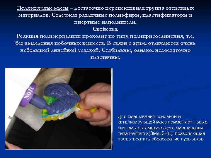 Polyester materials are a fairly promising group of impression materials. Contains various polyesters, plasticizers and excipients. Properties. The polymerization reaction proceeds according to the polyaddition type, i.e. without isolation by-substances... In this regard, they are characterized by a very small linear shrinkage. Stable, however, not plastic enough. New automatic mixing systems such as Pentamix (ЗМ / ESPE) are used to mix the base and catalytic mixes to prevent the formation of bubbles.
 Silicone impression materials The main advantages of Alphasil C-silicones: Working time varies depending on the amount of catalyst Low shrinkage percentage High accuracy and elasticity Impression shelf life - 1 week All materials are hydrophilic and thixotropic
 Disadvantages: - Not ideal quality when taking impressions with retraction threads - Requires careful manual mixing of masses and catalyst of dissimilar consistency - Difficulty of exact dosage of the catalyst, all "by eye" - Models cannot be cast on the impression repeatedly - Sensitivity to moisture - hygroscopicity - Low hydrophilicity - Insufficient adhesion to the tray - Possibility of toxic effect is described in the literature - No automatic mixing - Somewhat excessive stiffness of the base mass
 The main advantages of Betasil A-silicones: Easy mixing and precise dosage of the mass and catalyst due to the 1: 1 ratio Excellent hydrophilic and thixotropic properties of the masses Elasticity and tensile strength Perfect recovery forms after deformation Due to the high quality of the impression material, the impression can be used repeatedly. Thermoactive formula allows to adjust the hardening time of the material depending on the temperature. The total operating time varies from 2 to 4 minutes. Disadvantages: - Cannot be kneaded in latex gloves - A-silicones are slightly more expensive than C-silicones
 Thermoplastic masses soften at a temperature of 50 -70 ° C and become hard at an oral temperature (37 ° C). n Thermoplastics do not accurately represent the details of the prosthetic bed. The relief of the mucous membrane on the impression is displayed smoothed, since the mass has low fluidity. An accurate representation of the dentition cannot be obtained using a thermoplastic mass due to its hardening after cooling. Therefore, with the inclination of the teeth, pronounced equators of the teeth during removal, the impression is deformed.
 Thiodent is a completely non-shrinking material, which makes it possible to preserve impressions for a long time. The high elasticity of the impression material before the impression and the plasticity before vulcanization make it possible to obtain impressions that reflect the relief of the hard and soft tissues of the oral cavity. Cielast Advantages High elasticity of the impression. High impression accuracy. Good shape recovery after deformation. Several models can be cast on one print.

Using a plaster model, a spoon is made of wax, a small (up to 1 cm) wax handle is modeled in the area of \u200b\u200bthe front teeth, a model with a wax spoon is cast into a cuvette, the wax is melted, replaced with plastic, polymerized, processed, but the spoon is not polished.
You can make a spoon from self-hardening plastics (protacryl, carbodent, redont) by free molding and polymerization under pressure in water at room temperature. A plastic dough is prepared according to the previously described method, which is rolled out on a polyethylene plate with a glass rod to a thickness of 4 mm. From the obtained plate, a shape is cut out with a spatula, corresponding to the shape of the upper or lower toothless jaw. The resulting plate is placed on the model with the applied Isokola insulating layer. and molded.
The hardening of the plastic is accompanied by an exothermic reaction, which causes the plastic dough to drift slightly from the plaster model along the peripheral edge of the spoon. At this point, you must re-compress the edges of the spoon. In order to avoid deformation of the spoon, polymerization is recommended to be carried out in water at room temperature under pressure.
An individual spoon can be obtained from a standard AKR-P plate, which is softened in hot water and squeezed according to the model. In case of premature hardening, the unformed section of the plate is softened again and re-compressed according to the model. The excess plate is cut with scissors along the marked boundaries. From the leftovers
Fig. 181. Individual impression tray.
a - outer surface; b - inner surface; “- the moment of checking the boundaries of the spoon.

Rice. 182. Functional casts, edged with wax (solid black line).
a - blind man from the lower jaw; b - cast from the upper jaw.
the plates make a handle with a very hot spatula. An individual impression tray can be obtained from a plate of polystyrene or plexiglass with a thickness of up to 3 mm directly on a plaster model in a pneumatic press with a heater (PPS-1) and a dry-air polymerizer (PS-1).
Individual impression trays are fitted by the doctor in the patient's oral cavity, shortens the edges and forms them with a thermoplastic mass, using Herbst functional tests for this purpose.
After fitting the spoon, the doctor, depending on the flexibility and mobility of the mucous membrane of the prosthetic bed, takes a functional impression using elastic materials (thiodent, sielast), hardening (dentol, repin, gypsum) or thermoplastic masses (MCT-02, etc.).
After receiving an integral functional cast with plaster, it is edged. The edging is necessary to maintain the bulk of the prosthetic margin to ensure that the valve is closed during function. The edging is carried out as follows. With a chemical pencil, leaving 2-3 mm from the outer edge of the impression, mark a line along which a pre-prepared edging roller of wax 2-3 mm thick is attached with melted wax (Fig. 182).
Upon receipt of the model, the trace from the edging will maintain the outer boundaries of the neutral zone, which are necessary for the formation of the valve zone. The edging helps the dental technician to protect from violation of the border of the neutral zone when opening the plaster model, cast from a functional impression, which the doctor obtained using functional tests.