Is it possible to increase the ejection fraction of the heart. Heart emissions faction on ultrasound: how to determine the reasons for lowering the indicator
Before you put the patient a diagnosis of "chronic heart failure", the doctor conducts diagnostics with the mandatory definition of such an indicator as a fraction of emission. It reflects the amount of blood that the left ventricle pushes at the time of its reduction to the lumen of the aorta. That is, by such a study, it is possible to find out, the heart is effectively coping with its work or there is a need for prescribing heart medicines.
Code of Indicator FV
To evaluate the work of the heart, namely the left ventricle, the formulas of Teicholz or Simpson are used. It must be said that specifically of this department the blood gets into general blood circulation and at left-deuded deficiency is most often the clinical picture of heart failure is developing.

The closer this indicator is to normal, the better the main "engine" of the body and more favorable prediction for life and health is reduced. If the value obtained is much lacking up to normal, then it can be concluded that the internal organs do not receive the required amount of oxygen and nutrients With blood, which means the heart muscle must somehow support.
The calculation is made directly on the equipment on which the patient is examined. In modern ultrasound diagnostic cabinets, preference gives the Simpson method, which is considered more accurate, although the Takehold formula is not less common. The results of both methods may vary within 10%.
Ideally, the ejection fraction should be 50-60%. According to Simpson, the lower limit is 45%, and the Tayholz is 55%. Both methods differ enough high levels informative about the possibilities of myocardium to reduce. If the resulting value ranges in the range of 35-40%, they speak of launched heart failure. And more low indicators Frames are fraught with death.
Causes of decline of FV.
Low values \u200b\u200bcan be caused by pathologies as:
- Coronary artery disease . At the same time, blood flow by coronary arteries is reduced.
- Myocardial infarction in history. This leads to the replacement of the normal heart muscles by the scars that do not possess the necessary ability to reduce.
- Arrhythmia, Tachycardia and other ailments that violate the rhythm of the main "motor" of the body and conductivity.
- Cardiomyopathy. It consists in increasing or lengthening the muscles of the heart, which is due to a hormonal failure, long-term hypertension, heart defects.

Symptoms of the disease
The diagnosis of "reduced emission fraction" can be made on the basis of symptoms characteristic of this disease. Such patients often complain about attacks of shortness of breath, and both during physical exertion and at rest. Suddenly attacks can provoke a long walking, as well as the performance of the simplest work on the house: washing floors, cooking.
Often attacks arise in the night clock in the lying position. Loss of consciousness, weakness, fatigue and dizziness may mean that the brain and musculature of the skeleton experience blood deficit.
In the process of violation of blood circulation, a fluid delay occurs, which leads to the appearance of edema, and in severe cases they affect internal organs and tissues. A person begins to suffer from pain in the abdomen on the right side, and the stagnation of venous blood in the vessels of the liver can be fraught with cirrhosis.
These symptoms are characteristic to reduce the contractile function of the main "motor" of the body, but often it happens that the level of the ejection fraction is normal, so it is very important to be examined at least once a year and make echocardoscopy, especially with heart disease.
The increase in BV to 70-80% should also be alerted, as this may be a sign that the heart muscle cannot fill the growing heart failure and seeks to throw away in the aorta as a large blood concentration.
As the aless progressing, the performance of the LV will decline, and it is echocardioscopy in the dynamics that will allow you to catch this moment. High fraction Emission is characteristic of healthy people, in particular, athletes who have a heart muscle is quite traveled and is able to decline with more than that of an ordinary person, by force.
Treatment
You can increase the reduced FV. For this, doctors apply not only medicinal therapy, but also other methods:
- Prescribed drugs to improve the contractile ability of myocardium. These include cardiac glycosides, after which there is a noticeable improvement.
- To prevent overloading of the heart with an extra liquid, urge to comply with a diet with a restriction of the table salt up to 1.5 g per day and the resulting liquid to 1.5 liters per day. Along with this, the reception of diuretic drugs is prescribed.
- Appoint the reception of organoprotective agents that help protect the heart and vessels.
- Decide on the surgical operation. For example, valve prosthetics are carried out, shunts are installed on coronary vessels, etc. However, an extremely low ejection fraction may become a contraindication to the operation.
Prevention
Prevention to prevent the development of heart disease is of great importance, especially in children. In the age of high technology, when most of the work are performed by cars, as well as constantly deteriorating environmental Conditions Life and irregular nutrition The risk of heart diseases is raised at times.
Therefore, it is very important to eat right, play sports, more often to be in the fresh air. It is such a lifestyle that will ensure the normal reduction capacity of the heart and muscle training.
/ 30.07.2018
Low emission fraction. Exercises for the treatment of heart failure. Risk factors, symptoms.
Before you put the patient a diagnosis of "chronic heart failure", the doctor conducts diagnostics with the mandatory definition of such an indicator as a fraction of emission. It reflects the amount of blood that the left ventricle pushes at the time of its reduction to the lumen of the aorta. That is, by such a study, it is possible to find out, the heart is effectively coping with its work or there is a need for prescribing heart medicines.
The measurement principle is as follows: if the cardiac output is higher, then the cold returns to the place and becomes less diluted. And on the contrary, if the cardiac emission is low, it will take more time than the cold goes to the measurement place, and after that the cold will be more diluted. The calibration of the method was carried out simultaneous measurement using other methods. The method of measuring the thermodilation can be considered invasive to the extent that it requires the presence of a Swan-Ganz catheter on the right side of the heart and lungs.
However, this does not indicate cathetierization, and is mainly used where the catheter was introduced for other reasons, in particular to measure pressure. The accuracy of the method is not ideal, so several measurements are used sequentially, and the result is averaged.
Code of Indicator FV
To evaluate the work of the heart, namely the left ventricle, the formulas of Teicholz or Simpson are used. It must be said that specifically of this department the blood gets into general blood circulation and at left-deuded deficiency is most often the clinical picture of heart failure is developing.
Note: In this section, blood that enters the light pulmonary arteries will be called venous. Blood, which flows into pulmonary veins, and then into system artery, will be called arterial. The principle of the fic is a simple application of the law of conservation of matter.
When entering into relationships, we get. Thus, cardiac emission can be defined as. In this outcome, we used tributaries and outflows of oxygen amounts. Alternatively, we could use oxygen mass flows. Sometimes the volumetric flows of oxygen are also used. It is believed that this expression is the amount of oxygen that flows and follows from the blood if oxygen is in a gaseous state.
The closer this indicator is to normal, the better the main "engine" of the body and more favorable prediction for life and health is reduced. If the value obtained is much lacking up to normal, then we can conclude that the internal organs do not receive the necessary amount of oxygen and nutrients with blood, which means the heart muscle must somehow support.
Although this classic method is relatively accurate, it is rarely used for its invasiveness. The principle of the fic can be used for substances other than oxygen. This procedure avoids the need to collect arterial blood. Unfortunately, the method fails in the presence of weakly ventilated lung areas, which, of course, may in extreme cases go to pathological short circuits on the lungs.
This procedure can avoid the need for unpleasant central venous catheterization. The measurement occurs in such a way that the patient begins to breathe a mixture containing the substance. Then the partial pressure of this substance in arterial blood is measured. The advantage of this method is that when the gas is usually not present in the air, the venous inflow of this substance is zero before the measurement start.
The calculation is made directly on the equipment on which the patient is examined. In modern ultrasound diagnostic cabinets, preference gives the Simpson method, which is considered more accurate, although the Takehold formula is not less common. The results of both methods may vary within 10%.
Ideally, the ejection fraction should be 50-60%. According to Simpson, the lower limit is 45%, and the Tayholz is 55%. Both methods are characterized by a fairly high level of informative regarding myocardial opportunities to reduce. If the resulting value ranges in the range of 35-40%, they speak of launched heart failure. And even lower rates are fraught with mortal consequences.
And the heartbeat after treatment is calculated as. This method also uses the need for central venous catheterization. Summary. Non-invasive or low-invasive methods for measuring cardiac rhythm based on the use of the film principle can become accurate and inexpensive method Measurements of heart rate in the future. The potential use of oxygen and carbon dioxide has so far faced problems with the accuracy of transmission of partial pressures to concentrations in which it depends, for example, from the influence of the pH, the mutual interaction of both gases with hemoglobin, etc. Heterogeneity of the lungs can also cause problems.
Causes of decline of FV.
Low values \u200b\u200bcan be caused by pathologies as:
- Coronary artery disease. At the same time, blood flow by coronary arteries is reduced.
- Myocardial infarction in history. This leads to the replacement of the normal heart muscles by the scars that do not possess the necessary ability to reduce.
- Arrhythmia, Tachycardia and other ailments that violate the rhythm of the main "motor" of the body and conductivity.
- Cardiomyopathy. It consists in increasing or lengthening the muscles of the heart, which is due to a hormonal failure, long-term hypertension, heart defects.

Magnetic resonance: The resonant properties of protons in the kernel are changed at speeds. Magnetic resonance can be used as a precise method of measuring the stream of aorta. The road method, it is used only experimentally. Mathematical analysis of the pulse wave: the shape and amplitude of the pulse wave depend on heart Emission. The pulse wave is measured either using a classic air cuff or a sensor that sticks to the skin at the artery site. Therefore, mathematical analysis of this wave can be the meaning of cardiac output.
The problem is that the shape of the pulse wave also depends on the properties of the arteries. For example, in the elderly, where the elasticity of the aorta and its elastic effect is lost, systolic pressure is usually increasing, but the diastolic pressure remains normal. This method can be useful after calibration on a person using another method for constant monitoring of heart rate.
Symptoms of the disease
The diagnosis of "reduced emission fraction" can be made on the basis of symptoms characteristic of this disease. Such patients often complain about attacks of shortness of breath, and both during physical exertion and at rest. Suddenly attacks can provoke a long walking, as well as the performance of the simplest work on the house: washing floors, cooking.
Measurement of the impedance of the chest: electrical resistance chest You can measure several electrodes of chests. Resistance changes during the change of heart rate due to changes in the blood volume in the heart and therefore can be used to calculate the pulse frequency and the subsequent cardiac output. The method is cheap and non-invasive, but, unfortunately, inaccurate.
Acute myocardial ischemia left ventricular muscle fibers impairs the possibility of spasm and adherence. These changes can be reversible if ischemia does not last too long and does not end with ischemic fiber necrosis. IN last years He declared a number of observations indicating that ultimately the fate of muscle fibers covered in acute ischemia, the acute myocardial infarction was solved in several, maybe even a few hours after the appearance of the chest pain. Therefore, it is possible that the corresponding actions during this period - at least in some patients - to limit the amount of infarction of necrosis.
Often attacks arise in the night clock in the lying position. Loss of consciousness, weakness, fatigue and dizziness may mean that the brain and musculature of the skeleton experience blood deficit.
In the process of violation of blood circulation, a fluid delay occurs, which leads to the appearance of edema, and in severe cases they affect internal organs and tissues. A person begins to suffer from pain in the abdomen on the right side, and the stagnation of venous blood in the vessels of the liver can be fraught with cirrhosis.
Additional loads, increasing the need for oxygen necrosis of myocardium threatening increase within a heart attack, and can have an adverse effect on the patient's fate, even when their action is inconstant. When shrinking the fibers of a healthy area, covered with ischemia does not give a shrinkage, but rather under the influence of increasing pressure in the chamber of convexities acting as a kind of valve. The increase in the residual volume after the shrinkage, and the vulnerability of the left ventricle, due to its acute ischemia, leads to an increase in the pressure of the left ventricle, the end of the diastolic, and secondly, the increased pressure in the left atrium and the pulmonary veins inorganized to it exceeding the critical importance of this pressure predisposes To the formation of edema of the lungs, contrary to the expectation of both of these complications, does not always occur simultaneously: they saw in both cases. Isolated pulmonary edema and shock single cases. Simultaneous occurrence in the process of acute infarction of shock and edema of the lungs, as a rule, indicates very serious in harming the left ventricle and is subject to significantly higher mortality than any of these complications in an isolated form.
These symptoms are characteristic to reduce the contractile function of the main "motor" of the body, but often it happens that the level of the ejection fraction is normal, so it is very important to be examined at least once a year and make echocardoscopy, especially with heart disease.
The increase in BV to 70-80% should also be alerted, as this may be a sign that the heart muscle cannot fill the growing heart failure and seeks to throw away in the aorta as a large blood concentration.
If the hemodynamic consequences of the heart attack are developing in a less turbulent, they take the shape of a subacute or chronic left-sized deficiency, and in extreme cases - the so-called character. Low output heart syndrome. Last group Sometimes heavy shock is a descent of heart attacks in cases where the therapeutic intervention temporarily preserved patient's life, but not the restoration of normal blood circulation. The boundaries separating the above-mentioned clinical fluid syndromes, which is understandable to their overall pathogenesis.
As the aless progressing, the performance of the LV will decline, and it is echocardioscopy in the dynamics that will allow you to catch this moment. The high fraction of emissions is characteristic of healthy people, in particular, athletes who have a heart muscle is quite traveled and is able to decline with more than an ordinary person, force.
The hemodynamic monitoring section, the operation of the physiological compensatory mechanism, which makes an increase in the left ventricle causes the filling pressure - within certain limits - increase the impact volume. Insufficient supply of venous caused by absolute or relative hypovolemia can disrupt the operation of the mechanism. The only chance of improving in such cases, an increase in the reduction of the heart on the road of pharmacological or by improving the blood supply to the region affected by acute ischemia.
In patients with acute myocardial infarction hemodynamic equilibrium, often shaky. This balance can easily join the arrhythmic violated complications, it is dangerous to accelerate or slowing the danger of ventricular functions. These arrhythmias impede the functioning of compensating mechanisms that retain a threat of a minute, and even more threatening to increase the range of ischemic necrosis. The rapid and stable restoration of the optimal heart rhythm plays a decisive role in all cases when arithmetic and hemodynamic complications of the infarction coexist.
Treatment
You can increase the reduced FV. For this, doctors apply not only medicinal therapy, but also other methods:
- Prescribed drugs to improve the contractile ability of myocardium. These include cardiac glycosides, after which there is a noticeable improvement.
- To prevent overloading of the heart with an extra liquid, urge to comply with a diet with a restriction of the table salt up to 1.5 g per day and the resulting liquid to 1.5 liters per day. Along with this, the reception of diuretic drugs is prescribed.
- Appoint the reception of organoprotective agents that help protect the heart and vessels.
- Decide on the surgical operation. For example, they are carried out, shunts are installed on coronary vessels, etc. However, an extremely low emission fraction may become a contraindication to the operation.
Prevention
Prevention to prevent the development of heart disease is of great importance, especially in children. In the age of high technologies, when most of the work are performed by cars, as well as constantly deteriorating environmental conditions and irregular nutrition, the risk of developing heart disease rises at times.
This is usually a prerequisite successful treatment Hemodynamic complications. The elimination of these additional factors plays an important role in the prevention of hemodynamic complications of a heart attack, as well as in the treatment of already developed complications. Late reception of hemodynamic complications usually indicates a heart attack or a complication of a mechanical type. The diagnosis and treatment of acute pulmonary swelling, complicating the recent myocardial infarction, are based on the principles set forth in Ch. The improvement achieved in mechanical ventilation should be used for high-speed drugs and furosemide preparations.
Therefore, it is very important to eat right, play sports, more often to be in the fresh air. It is such a lifestyle that will ensure the normal reduction capacity of the heart and muscle training.
During medical examinations, many patients often hear incomprehensible concepts and diagnoses. When a person has problems with the heart muscle, qualified specialists can calculate the effectiveness of cardiac activity. During the reduction of the heart muscle, blood pumping occurs, and the ejection fraction is the amount of blood plasma that enters the vessels. Experts measure this process in percent.
The introduction of morphine in the hope of monitoring the edema of the lungs in spontaneous patients with breathing, contraindicated for the reasons set forth on page 3. Stroke is even more than 50% of mortality at intensive therapy. Universal consensus was not achieved relative to the optimal method of treating these patients pharmacological treatment, although in recent years a lot of information has appeared on this issue. The direct goal of treatment is an increase in the volume of the volume of the left ventricle's ejection to cover the requirements for metabolic tissue.
Most often in order to measure the amount of blood, the doctors spend measurements on the left ventricle. Since blood is moving from it to a large circulation of blood circulation. If there is a reduced level of fraction of the emission of the left ventricle of a person, this can contribute to the appearance of heart failure.
Therefore, it is recommended to regularly access a qualified technician for diagnostics. In order to explore this process, you can use several methods. The simplest of them is an ultrasound study. He is pretty good because the doctor can find out how active and effective are the reductions of the heart muscle. This method is quite simple and convenient, and also does not provoke the appearance of side effects and is not dangerous for the human body.
Patients whose pressure of the left ventricle is only moderately increased, often reach this goal, additionally increasing the filling pressure by quickly intravenous infusion of low molecular weight dextran. In terms of balance, 4 - intense oxygen therapy 49 is the most economical way to increase the volume of emissions; The increase in the volume of ejection obtained on this path increases the need for myocardial oxygen to a much lesser extent than similar growth with cardiac abbreviations.
Suitable only for patients with strong shock syndrome, which have no symptoms of pulmonary edema. In patients with hemodynamic observation, the decision to aim on the use of dextran can be measured in diastolic arterial pressure. In patients with the right to treatment with low molecular weight dextran, we consider this drug the first choice in the fight against shock, which is associated with a recent myocardial infarction. At the same time, the dextran infusion, the patient should obtain approximately 90 mg eq trisamine to compensate for the accompanying metabolic acidosis.
The second method of diagnostics is isotopic ventriculography. During the use of this method, you can find out how efficiency is a fraction of emissions from the right and left ventricles. This option is more expensive, so quite often patients are diagnosed with ultrasound research.

In order to make any conclusions, it is necessary to know what the norm of the ejection fraction in the person. After the diagnosis was carried out, it is imperative to compare with the norm, and then the doctor must sum up and appoint a correct and effective course of treatment. If the ejection fraction of the cardiac muscle is normal and at the same time a person does not feel any noticeable violations in the work of the heart, it means everything is fine. The norm of this indicator is 55-70 percent. Even if a person is in a calm state, his left ventricle can throw in the vessels more than half of the blood, which is in it.
If there is a low fraction of the emission fraction in humans, a qualified specialist should send it to the necessary additional studies in order to determine the cause of this process. Frequently often factor of a reduced ejection fraction may assume the development of various heart diseases, such as cardiac insufficiency. It can appear due to the vices of the heart muscle, as well as ischemic illness. All these diseases are rather dangerous to human life, so they must be found as quickly as possible and begin an effective and effective treatment.
If problems and deviations from the norm of the heart emission faction are observed, it is necessary to refer to a qualified specialist who will conduct diagnostics. After diagnostic measures, the doctor must learn the reason for the appearance of this defect. Then, the doctor must appoint proper and effective treatment in order to prevent the symptoms and signs of the heart disease. The main feature of the prevention of the disease is the permanent control of the doctor and comply with all its recommendations. In order to protect their health, it is necessary at the very first symptoms to refer to a qualified doctor for diagnostics.
website - Medical portal about heart and vessels. Here you will find information about the reasons, clinical manifestations, diagnosis, traditional and folk methods Treating cardiological diseases of adults and children. And also on how to keep the heart is healthy, and the vessels are clean to the most advanced years.
Do not use the information posted on the site without prior consultation with your doctor!
Site authors - practitioners practitioners. Each article is the concentrate of their personal experience and knowledge, honored for years of study at the university received from colleagues and in the process of postgraduate training. They not only share unique information in articles, but also conduct virtual reception - answer the questions you specify in the comments give recommendations, help to understand the results of surveys and appointments.
Everything, even very difficult to understand the topic, are presented in a simple, understandable language and are designed for readers without medical training. For your convenience, all the themes are divided into categories.
Arrhythmia
According to the World Health Organization, arrhythmias - more than 40% of people over 50 years old, suffer from violations of heart rhythm. However, not only they. This cunning ailment detects even in children and is often in the first second year of life. What is he cunning? And what is sometimes masks under the diseases of the heart of the pathology of other vital organs. Another unpleasant feature of arrhythmia is a secretiveness: while the disease does not go too far, you can not guess about it ...
- how to identify arrhythmia at an early stage;
- what kind of forms are most dangerous and why;
- when the patient is enough, and in what cases can not do without surgery;
- how and how much do you live with arrhythmias;
- what attacks of the rhythm disorders require immediate challenge the ambulance, and at what it is enough to take the pill of the sedative.
And also all about the symptoms, prevention, diagnosis and treatment of various types of arrhythmias.
Atherosclerosis
The main role in the development of atherosclerosis plays an excess of cholesterol in food, they write in all newspapers, but why then in families where everyone feeds is the same, only someone is often sick? Atherosclerosis is known for more than a century, but much in its nature remained unsolvable. Does it despair? Of course not! Specialists Site tell what success in the fight against this ailment has reached modern medicineHow to prevent it and how to treat it effectively.

- why margarine is harmful to cream oil for people with vascular damage;
- and what he is dangerous;
- why do not help non-baldterine diets;
- from what will have to abandon the whole life sick with;
- how to avoid and preserve the clarity of the mind to a deep old age.
Heart diseases
In addition to angina, hypertension, myocardial infarction and congenital defects Hearts There are a lot of other cardiac ailments, which many never heard. Do you know, for example, what is not only a planet, but also a diagnosis? Or that in the heart muscle can grow a tumor? About these and other diseases of the heart of adults and children tells the same category.
- and how to render emergency help patient in this state;
- what and what to do for the first to do not move into the second;
- why the heart of alcoholics increases in size;
- what is dangerous prolaks mitral valve;
- for what symptoms can be suspected of your child's heart disease;
- what cardiological ailments are more threatened to women, and what are men.

Diseases of Vessels
The vessels permeate the whole body of man, so the symptoms of their defeat are very and very diverse. Many vascular alaughs at first are little bothering the patient, but lead to formidable complications, disability and even death. Can a person without medical education to identify the pathology of the vessels? Of course, yes, if they know their clinical manifestations, which this rubric will tell.
In addition, it contains information:
- about medical preparations and folk remedies for the treatment of vessels;
- about what doctor to contact with suspected vascular problems;
- what vascular pathologies are mortally dangerous;
- from which the veins are swept;
- how to keep the health of veins and arteries for life.
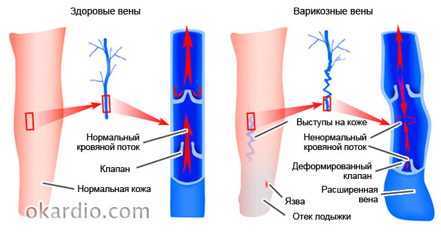
Varicose
Varicose veins (varicose veins) - a disease in which the lumens of some veins (feet, esophagus, rectum, etc.) become too wide, which leads to a violation of blood flow in the affected organ or body part. In the launched cases, this ailment is heal with great difficulty, but in the first stage it is quite possible to curb. How to do this, read in the "Varicose" rubric.
 Click on photo to enlarge
 Also from it you will learn:
- what are the ointments for the treatment of varicose varicose and which is more effective;
- why some patients with varicose veins of the lower extremities doctors prohibit running;
- and to whom it threatens;
- how to strengthen veins by folk remedies;
- how to avoid the formation of blood clots in the affected veins.
Pressure
- So widespread understood that many consider it ... a normal state. From here and statistics: only 9% of people suffering from high pressure hold it under control. And 20% of hypertensive and consider themselves healthy at all, since their disease proceeds asymptomatic. But the risk of getting a heart attack or stroke is not less from this! Although less dangerous than high, but also gives a lot of problems and threatens with serious complications.

In addition, you will learn:
- how to "deceive" heredity, if both parents suffered hypertension;
- how to help yourself and close in hypertensive crisis;
- why increases pressure in young age;
- how to keep pressure under control without medication, using therapeutic herbs and certain products.
Diagnostics
In the heading devoted to the diagnosis of heart disease and vessels, articles are collected about the types of surveys who pass cardiological patients. As well as testimony and contraindications to them, interpretation of results, efficiency and procedure for conducting procedures.
You also find answers to questions:
- what types of diagnostic research should even pass healthy people;
- why appoint angiography to those who suffered a myocardial infarction and stroke;

Stroke
Stroke (acute violation of cerebral circulation) is consistently among the top ten dangerous diseases. People over 55 years old, hypertensive, smokers and those who suffer from depression have become the greatest risk of its development. It turns out that optimism and good nature reduce the risk of strokes almost 2 times! But there are other factors that effectively help him avoid.
The heading dedicated to the strokes tells about the reasons, types, symptoms and treatment of this insidious disease. And also - about rehabilitation measures that help restore lost functions to those who have soles.

In addition, you will learn from here:
- about the difference clinical manifestations strokes in men and women;
- about what is a pre-primary state;
- about folk remedies for the treatment of consequences of strokes;
- on the modern methods of rapid recovery after the transferred stroke.
Infarction
Myocardial infarction is considered to be the disease of the elderly men. But he is still the greatest danger, it is still not for them, but for people of working age and women over 75 years old. It is in these groups that the death rates are the highest. However, it is not worth relaxing to anyone: today, heart attacks overtake even young, sports and healthy. More precisely, the sustained.
In the heading "InfarTr", experts tell about everything that it is important to know everyone who wants to avoid this ailment. And those who have already suffered a myocardial infarction will find a lot of useful tips on treatment and rehabilitation.

- about what diseases are sometimes masked by heart attack;
- how to provide urgent help with acute pain in the field of heart;
- about differences in the clinic and the flow of myocardial infarction in men and women;
- about the anti-infarction diet and safe for the heart of life;
- about why a sick infarction must be delivered to a doctor for 90 minutes.
Pulsa Disorders
Speaking of violations of the pulse, we usually mean its frequency. However, the doctor assesses not only the patient's heartbeat speed, but also other pulse wave records: rhythm, filling, voltage, form ... Roman Galen surgeon at one time described the whole 27 of its characteristics!

The change in individual pulse parameters reflects not only the heart and blood vessels, but also other organism systems, for example, endocrine. Want to know more about it? Read the heading materials.
Here you will find answers to questions:
- why, with complaints about the violation of the pulse, you may be sent to the thyroid examination;
- can the slowdown of heart rate (bradycardia) cause the heart stop;
- what does it mean and what it is dangerous;
- as interconnected pulse frequency and fat burning rate when weight loss.
Operations
Many diseases of the heart and vessels, which for another 20-30 years ago, ordered people on life disability, today are successfully cured. As a rule, surgically. Modern cardiac surgery saves even those who have recently left no chance of life. And most of the operations are held now through tiny punctures, not cuts, as before. This not only gives a high cosmetic effect, but it is much easier to transfer. And also reduces the time of postoperative rehabilitation several times.
In the heading "Operations" you will find materials on surgical methods for the treatment of varicose veins, shunting of vessels, installation of intravascular stents, cordial valve prosthetics and much more.
And also learn:
- what technique does not leave scars;
- as operations on the heart and vessels affect the quality of life of the patient;
- what is the differences in operations and vessels;
- under what diseases are conducted and what is the duration healthy Life after him;
- what is better in heart disease - to be treated with pills and injections or make an operation.

Rest
The "rest" entered the materials that do not correspond to the subject of other headings of the site. It contains information about rare cardiological ailments, about myths, misconceptions and interesting facts related to the health of the heart, about the incomprehensible symptoms of their meaning, about the achievements of modern cardiology and many other things.
- on the provision of first aid to oneself and other in various urgent states;
- about the child;
- about sharp bleeding and methods of their stop;
- o and food habits;
- about folk methods of strengthening and improving cardiovascular vascular system.

Preparations
"Preparations" - perhaps the most important heading of the site. After all, the most valuable information about the disease - how to treat it. We do not give magic recipes for the cure of heavy ailments with one tablet, we honestly and truthfully tell about the preparations, everything is as it is. What they are good and the bad things are shown and contraindicated, what differ from the analogs and how they affect the body. These are not calls for self-treatment, it is necessary so that you are well owned by "weapons", which you have to fight with the disease.

Here you will find:
- reviews and comparison of groups of drugs;
- information on what can be taken without the appointment of a doctor, and that in no case cannot be;
- list of grounds for selecting a means;
- information about cheap analogues of expensive imported drugs;
- data on the side effects of cardiac drugs that manufacturers are silent.
And many more important, useful and valuable, which will make you healthier, stronger and happier!
Let your heart and vessels always be healthy!
What is an emission fraction and why need to be evaluated?
The ejection fraction (FV) is an indicator reflecting the volume of blood pushed by the left ventricle (LV) at the time of its reduction (systole) into the lumen of the aorta. FV is calculated on the basis of the ratio of blood volume emitted in the aorta, to the blood volume located in the left ventricle at the time of its relaxation (diastole). That is, when the ventricle is relaxed, it contains blood from the left atrium (the final diastolic volume - KDO), and then, shrinking, it pushes a part of the blood into the lumen of the aorta. This is the part of blood and is a fraction of emission expressed as a percentage.
The blood release fraction is the magnitude that is technically just to calculate, and which has a sufficiently high informativeness relative to the contractile ability of myocardium. This value largely depends on the need to prescribe heartbreaks, and the forecast for patients with cardiovascular failure is determined.
The closer to the normal values \u200b\u200bof the LV Emission Fraction in the patient, the better its heart is reduced and a favorable forecast for life and health. If the emission fraction is much lower than the norm, it means that the heart cannot be normally reduced and ensure the whole organism with blood, And in this case, the heart muscle should be supported by medicines.
How do the emission fraction calculate?

This indicator can be calculated using the Takehold or Simpson formula. The calculation is carried out using a program that automatically calculates the result depending on the final systolic and diastolic volume of the left ventricle, as well as its size.
More successful is the calculation according to the Simpson method, Since the Takeholder in the section of the study with two-dimensional echo-kg may not be hit by small areas of myocardium with a disturbed local shortcutivity, while with the method of simpson in the circumference section, more significant portions of myocardium fall into the section.
Despite the fact that the Outdated Equipment uses the Tayholz method, modern bubbies of ultrasonic diagnostics prefer to evaluate the ejection fraction by the Simpson method. The results obtained, by the way, may differ - depending on the method by magnitude within 10%.
Normal indicators of FV.
The normal value of the emission fraction is different from different peopleand also depends on the equipment on which the study is carried out, and from the method according to which the fraction is calculated.
The averaged values \u200b\u200bare approximately 50-60% The lower boundary of the norm according to the Simpson formula is at least 45%, according to the Takehold formula, at least 55%. This percentage means that this amount of blood for one heart abbreviation is necessary to push the heart into the lumen of the aorta to ensure adequate delivery of oxygen to the internal organs.
About launched heart failure say 35-40% , even lower values \u200b\u200bare fraught with configurations.
In childrenin the period of newborn, FV is at least 60%, mainly 60-80%, gradually reaching the usual indicators of the norm as they grow.

Of the abnormalities from the norm, more often than an increased fraction of emissions, there is a decrease in its value due to various diseases.
If the indicator is reduced, it means that the heart muscle can not be reduced enough, As a result, the volume of the beamped blood decreases, and the internal organs, and, first of all, the brain, is obtained less oxygen.
Sometimes in the conclusion of echocardioscopy, it can be seen that the value of FV is above average indicators (60% or more). As a rule, in such cases, the indicator is not more than 80%, since the larger blood volume of the left ventricle due to physiological features can not be expelled in the aorta.
As a rule, high FV is observed in healthy persons in the absence of other cardiological pathology, As well as athletes with the training cardiac muscle, when the heart is reduced with a greater force at every blow than an ordinary person, and the larger percentage of blood contained in it is expelled in the aorta.
In addition, in case the patient has a LV as a manifestation of hypertrophic cardiomyopathy or arterial hypertension, an increased FV may indicate that with the bridal muscle can still compensate for beginner heart failure And seeks to drive out in the aorta as much as possible blood. With progression of heart failure, FV is gradually decreasing, therefore, for patients with clinically manifested CHF, it is very important to perform echocardioscopy in the dynamics in order not to miss a decrease in FV.
Causes of a reduced heart emission fraction
The main cause of the disturbance of the systolic (contractile) function of myocardial is the development (CHF). In their own, queue, CXN arises and progresses due to diseases such as:

The most common cause of a reduction in heart emissions are acute or transferred myocardial infarction, accompanied by a decrease in the global or local reduction in the myocardial of the left ventricle.
Symptoms of reduced emission fraction
All the symptoms that can be suspected are due to CXN. Therefore, the symptoms of this disease goes first.
However, according to observations of practicing doctors of ultrasound diagnostics, the following is often observed - in patients with pronounced signs of the CHF, the emission fraction indicator remains within the normal limits, while in persons with missing explicit symptoms, the emission fraction indicator is significantly reduced. Therefore, despite the absence of symptoms, patients with the presence of cardiac pathology necessarily at least once a year to perform echocardoscopy.

So, to symptoms, allowing to suspect a disruption of myocardial cuts, belong:
- Attacks of shortness of shortness of breath or at exercise, as well as in the lying position, especially at night,
- The load provoking the occurrence may be different - from significant, for example, walking for long distances (more than 500-1000m), to minimal household activity, when the patient is hard to perform the simplest manipulations - cooking, laundry starting, walking to the next room and t . D,
- Weakness, fatigue, dizziness, sometimes loss of consciousness - all this indicates that skeletal muscles and the brain receive little blood,
In the absence of competent treatment of myocardial systolic dysfunction, such symptoms progress, grow and are increasingly transferred to the patient, so if you even have one of them, you should obtain a consultation of a doctor or cardiologist.
In which cases requires a reduced emission fraction?
Of course, no doctor will offer you to pass a low indicator obtained by heart ultrasound. At first, the doctor must reveal the reason for the reduced FV, And then appoint treatment of a causal disease. Depending on it, treatment may differ, for example, the reception of nitroglycerin drugs for ischemic disease, surgical correction Heart defects, hypotensive drugs in hypertension, etc. Patient It is important to understand that if there is a decrease in the ejection fraction, it means that heart failure really develops and it is necessary to fulfill the recommendations of the doctor for a long time and scarpurose.

How to increase the reduced emission fraction?
In addition to drugs affecting the causal disease, medications are prescribed to the patient, capable of improving myocardial contractility. These include (Digoxin, Stroofantin, Corgalicon). However, they are appointed a strictly attending physician and independent uncontrolled application is unacceptableSince poisoning can occur - glycoside intoxication.

To prevent heart overload volume, That is, excessive liquid, shows the observance of the diet with the restriction of the table salt up to 1.5 gr per day and with the restriction of the injubiled fluid to 1.5 l per day. Also successfully used - diakarb, diouver, Verosampirov, Indapamide, Toramsemed, etc.
To protect the heart and vessels from the inside Preparations with so-called organoprotective properties are used - ACE inhibitors. These include Enalapril (ENAP, ENAM), Perindopril (Preshaur, Prestentes), Lisinopil, Captopril (Kopoten). Also from drugs with similar properties, inhibitors of Ara II - Losartan (Lorist, Lozart), Valsartan (Valz) and others are widespread.
The treatment diagram is always selected individually, But the patient should be ready for the fact that the ejection fraction is not normalized immediately, and the symptoms can disturb some time after the start of therapy.
However, in the case of severe heart failure (III-IV functional class) with an extremely low emission fraction, the operation may be contraindicated. For example, contraindication The prosthetics of the mitral valve is the decrease in the FV is less than 20%, and to the implantation of the pacemaker - less than 35%. However, contraindications to operations are detected at full-time examination of the cardiac surgeon.
Prevention

The prophylactic orientation of the on the low fraction of the emission remains particularly relevant in a modern environmentally unfavorable setting, in the era of a low-fat lifestyle behind computers and nutrition by low-oats.
Even on the basis of this, it can be said that fresh holidays outside the city in the fresh air, healthy nutrition, adequate physical exertion (walking, easy run, charging, gymnastics), rejection of bad habits - all this is the key to long and proper functioning of cardio-vascular system with normal contractile ability and cordial muscle training.
Video: Lecture "Heart failure with a stored emission fraction - clinical dilemma"
Patients who received the direction for medical diagnostics of the heart and blood vessels, meet such a concept as a fraction of emission. It is measured during ultrasound, contrasting x-ray and echoc.
In this article, the reader will get acquainted with the definition of "Heart Emission", norms and decoding, and also learns about the methods of treatment and prevention.
For all issues, you can contact the portal specialists.
Competent consultations are carried out free of charge 24 hours a day.
Concepts and symptoms
The emission fraction is an indicator that determines the effectiveness of the health of the heart of the heart body at the moment of impact. It is measured as a percentage ratio of blood volume falling into the vessels in the state of the ventricle systole. For example, with 100 ml in the vascular system, 65 ml falls, thus, the heart emission will be equal to 65%.
Mainly, the measurement is carried out left ventricle, since it enters blood circulation from it big circle. If there is a lack of blood in this ventricle, it becomes the cause of heart failure, which entails the development of diseases of the authority.
The emission fraction is appointed not to all patients, but only those who complain about:
- pain syndrome in the chest;
- systematic interruptions in the body;
- tachycardia;
- dyspnea;
- frequent dizziness and fainting;
- swelling of the lower extremities;
- fast fatigue and weakness;
- reduced productivity.

As a rule, the first study is an electrocardiogram and ultrasound. These surveys allow you to find out to which extent is the cardiac emission of both the left ventricle and the right. Diagnosis is distinguished by low price, high informativeness, and there is no specific preparation. The availability of the procedure is due to the fact that any ultrasound equipment can provide data on the fraction.
Normal fraction emission
Human heart, even without external stimuli, continues to work, pushing over 50% of blood at every systolic condition. If this indicator begins to decrease to the plank less than 50%, insufficiency is diagnosed. As a result, the reduction in the volume is developing myocardium, ischemia, vice, etc.
The emission fraction varies in the range of 55-70 percent is the norm. The decline to the mark of 35-40 percent entails dangerous interruptions. To prevent the fatal drop, you need, at least once a year to visit the cardiologist. Persons who have reached the age of 40 are a mandatory procedure. The symptomatic picture described above is a faithful reason to appeal to a qualified cardiologist.
Cardiac release when diagnosing a patient's body with pathologies in a cardiovascular system, an important priority is to determine the individual minimum threshold. Based on information, the doctor may diagnose and assign correct therapy.
Ultrasound - norms and decoding
At the end ultrasound examinationThe diagnost is a protocol, which makes all the obtained data on the state of the left ventricle. Subsequently, information is subjected to decoding. When detecting pathologies, the doctor explains the results obtained and the diagnosis is established.
Not even possessing medical educationThe person can independently decipher the main indicators and see the clinical picture of the investigated body. The decoding occurs by the method of comparing the received information with the norm table.
- emission fraction, gap: 55 -60%;
- the size of the atrium of the right chamber: 2.7-4.5 cm;
- impact volume: 60-100 ml;
- aortic diameter: 2.1-4.1 cm;
- the thickness of the diastolic wall: 0.75-1.1 cm;
- systole size: 3.1-4.3 cm;
- the atrium size of the left chamber: 1.9 to 4 cm.
The above indicators must be considered in the aggregate volume. The deviation from the norm of one is not suspicion of the pathological process, but may require additional diagnostics.
On the portal you can download for free:
How to treat low level of fraction?
With information about the rate of cardiac output, the reader may analyze the health of the body. When the release of the left ventricle turns out to be lower than the norm, it is recommended to take advice from the cardiologist in a short time. It is worth noting that the doctors are primarily interested in not the presence of pathology, but the reason for the development of alend. Therefore, after ultrasound, additional research is often carried out.
Low ventricular emission is most often characterized by poor well-being, swelling and shortness of breath. How to increase the volume of the fraction? We live in the era of progressive medicine, so in the arsenal of doctors to increase blood emissions in the vessels, therapy occupies the first place. Basically, the ward is located on an outpatient treatment, during which experts are watching the activities of the heart and system of vessels. In addition to drug treatment, surgical intervention is sometimes produced.
- the reception of the liquid is strictly regulated and is 1.5-2 liters of water per day;
- failure to salt, seasonings and dishes containing it;
- dietary diet;
- moderate physical activity;
- reception of drugs: urinary tract stimulants, inhibitors, adrenaline blockers, digoxin, etc.
Restore blood supply can be surgical method. As a rule, operations are prescribed to patients who diagnosed severe heart rate or valve apparatus. Often, valves are subjected to resection and set prostheses. This approach allows to normalize heart rate, eliminate arrhythmia and fibrillation. It should be noted that the operations are carried out if there is a high danger to a person's life. In all other cases, therapy is carried out.
Preventive methods
With absence genetic predisposition The fraction can be kept normal to diseases of the cardiovascular system.
- daily charging;
- consumption of products saturated with iron;
- do not abuse alcohol-containing drinks and tobacco;
- observe a healthy mode;
- 2-3 times a week to make aerobics;
- choose dishes with low salt.
The main key is aerobics. There is such an opinion that in diseases of the heart and vessels, physical exertion is harmful. It is a myth.
Damage can only bring weight lifting, i.e. gym For patients are prohibited. Aerobic exercises on the contrary strengthen the walls of the vessels and do not overload the heart. Such classes improve muscle functions, deriving oxygen from the blood. It is necessary to engage gradually increasing the load.
According to statistics of the 20th century, the people of old age were most often affected by the cordially vascular diseases. To date, it affected the younger generation. The main risk group includes residents of metropolis, which suffer from low levels of clean air and exhaust gases. Therefore, it is very important to every person every year a medical examination is not only at the cardiologist, but also other doctors. Remember that only you are responsible for your health!
cardiologya.com.
If you have already passed ultrasound procedure Kidney or, for example, the abdominal organs, then you remember that for approximate decryptions of their results, it is most often not necessary to contact the doctor - the basic information can be found before visiting the doctor, with an independent reading of the conclusion. Heart ultrasound results are not so easy for understanding, therefore it is not easy to solve them, especially if you disassemble each tippet.
You can, of course, simply look at the last lines of the form, where the general summary is written in the study, but this also does not always clarify the situation. So that you can easily understand the results obtained, we give the main norms of heart ultrasound and possible pathological changes that can be installed by this method.
Norms in ultrasound for heart chambers
To begin with, we give a few digits that are necessarily found in each conclusion of Doppler Echocardiography. They reflect different parameters of the structure and the functions of individual heart chambers. If you are a pedant, and responsible approach to decipher your data, pay the maximum attention to this section. Perhaps here you will meet the most detailed information, in comparison with other Internet sources designed for a wide range of readers. In different sources, data can be somewhat different; Here are the numbers based on the materials of the "Norma in Medicine" manual (Moscow, 2001).
Left ventricular parameters
Light ventricle myocardium mass: Men - 135-182, women - 95-141
Light ventricle myocardial mass index (in the form is often designated as immulzh): Men 71-94 g / m2, women 71-89 g / m2.
Of course-diastolic volume (KDO) of the left ventricle (the volume of the ventricle, which he has at rest): Men - 112 ± 27 (65-193) ml, women 89 ± 20 (59-136) ml
Of course-diastolic size (CDR) left ventricle (The size of the ventricle in centimeters, which he has at rest): 4.6 - 5.7 cm
Eltimate Systolic Size (CER) left ventricle (The size of the ventricle that it has during the reduction): 3.1 - 4.3 cm
Wall thickness in diastole (outside the heart cuts): 1.1 cm
With hypertrophy - increasing the thickness of the ventricular wall, due to too much load on the heart - this indicator increases. Figures 1.2 - 1.4 cm talk about minor hypertrophy, 1.4-1.6 - about the average, 1.6-2.0 - about considerable, and the value more than 2 cm testifies to high degree hypertrophy.
Emission fraction (FV): 55-60%.
In a state of rest of the ventricle is filled with blood, which is not completely thrown out of them during abbreviations (systole). The emission fraction shows what amount of blood relative to its total amount eats the heart with each reduction, it is normal a little longer than half. With a decrease in the indicator of FV, they talk about heart failure, this means that the body is ineffectively pumping blood, and it can be stated.
Impact volume (The amount of blood, which is ejected by the left ventricle for one reduction): 60-100 ml.
Parameters of right ventricle
Wall thickness: 5 ml
0.75-1.25 cm / m2 size index / m2
Diastolic size (size in peace) 0.95-2.05 cm
The parameters of the interventricular partition
Thickness at rest (diastolic thickness): 0.75-1.1 cm
Excursion (moving from side to side during heart cuts): 0.5-0.95 cm. The increase in this indicator is observed, for example, with some heart defects.
Parameters of the right atrium
For this heart chamber, only the value of the QDO is determined - the volume is at rest. A value of less than 20 ml indicates a decrease in CDO, the indicator greater than 100 ml indicates its increase, and the KDO is more than 300 ml with a very significant increase in the right atrium.
Parameters of the left atrium
Size: 1.85-3.3 cm
Size index: 1.45 - 2.9 cm / m2.
Most likely, even a very detailed study of the parameters of cardiac cameras will not give you particularly clear answers to the question of the state of your health. You can simply compare your performance with optimal and on this basis to make preliminary conclusions about whether you are generally normal. For more information, refer to a specialist; For its wider lighting, the volume of this article is too small.
Norms in ultrasound for heart valves
With regard to decryption of valve surveys, it should be a simpler task. You will be enough to look at the general conclusion about their condition. The main, most frequent pathological processes are only two: it is stenosis and valve failure.
Term "stenosis" The narrowing of the hole of the valve is denoted by the above the underlying camera of the heart with difficulty pumps blood through it and can be undergoing hypertrophy, which we talked about in the previous section.
Failure - This is the opposite state. If the valve sash is, in the norm, preventing the reverse current of blood, for some reason they cease to perform their functions, the blood passed from one heart chamber to another, partially returns back, reducing the efficiency of the organ.
Depending on the severity of disorders, stenosis and insufficiency can be 1.2 or 3 degrees. The higher the degree, the more serious pathology.
Sometimes in conclusion, the ultrasound of the heart can be found such a definition as "relative failure." At this state, the valve itself remains normal, and blood flow disorders arise due to the fact that pathological changes occur in adjacent heart chambers.
Standards in ultrasound for pericardia
Pericardi, or a near-handing bag - this is a "bag" that surrounds the heart outside. It grows with an organ in the field of vessels, in its upper part, and between him and the heart itself there is a sliding cavity.
The most frequent pathology of Pericarda is inflammatory process, or pericarditis. With the pericardia between the window, the spikes can be formed and the liquid can be accumulated. Normally, it is 10-30 ml, 100 ml speaks of a small accumulation, and over 500 - about a significant accumulation of fluid, which can solve the completeness of the full work of the heart and squeezing ...
To master the specialty of the cardiologist, the person must first study at the university for 6 years, and then for a minimum of the year to separately study cardiology. A qualified doctor has all the necessary knowledge, thanks to which he can not only easily decipher the conclusion to the ultrasound of the heart, but also to diagnose on its basis and prescribe treatment. For this reason, deciphering the results of such a complex study, as an echo cardiography, should be provided with a profile specialist, and not try to do it yourself, long and unsuccessfully "picking up" in numbers and trying to understand what they mean certain indicators. This will save you a lot of time and nerves, as you do not have to worry about your own, probably disappointing and, even more likely, the wrong conclusions about the state of your health.
analizi-uzi.com.
Code of Indicator FV
To evaluate the work of the heart, namely the left ventricle, the formulas of Teicholz or Simpson are used. It must be said that specifically of this department the blood gets into general blood circulation and at left-deuded deficiency is most often the clinical picture of heart failure is developing.

The closer this indicator is to normal, the better the main "engine" of the body and more favorable prediction for life and health is reduced. If the value obtained is much lacking up to normal, then we can conclude that the internal organs do not receive the necessary amount of oxygen and nutrients with blood, which means the heart muscle must somehow support.
The calculation is made directly on the equipment on which the patient is examined. In modern ultrasound diagnostic cabinets, preference gives the Simpson method, which is considered more accurate, although the Takehold formula is not less common. The results of both methods may vary within 10%.
Ideally, the ejection fraction should be 50-60%. According to Simpson, the lower limit is 45%, and the Tayholz is 55%. Both methods are characterized by a fairly high level of informative regarding myocardial opportunities to reduce. If the resulting value ranges in the range of 35-40%, they speak of launched heart failure. And even lower rates are fraught with mortal consequences.
Causes of decline of FV.
Low values \u200b\u200bcan be caused by pathologies as:
- Coronary artery disease. At the same time, blood flow by coronary arteries is reduced.
- Myocardial infarction in history. This leads to the replacement of the normal heart muscles by the scars that do not possess the necessary ability to reduce.
- Arrhythmia, Tachycardia and other ailments that violate the rhythm of the main "motor" of the body and conductivity.
- Cardiomyopathy. It consists in increasing or lengthening the muscles of the heart, which is due to a hormonal failure, long-term hypertension, heart defects.

Symptoms of the disease
The diagnosis of "reduced emission fraction" can be made on the basis of symptoms characteristic of this disease. Such patients often complain about attacks of shortness of breath, and both during physical exertion and at rest. Suddenly attacks can provoke a long walking, as well as the performance of the simplest work on the house: washing floors, cooking.
In the process of violation of blood circulation, a fluid delay occurs, which leads to the appearance of edema, and in severe cases they affect internal organs and tissues. A person begins to suffer from pain in the abdomen on the right side, and the stagnation of venous blood in the vessels of the liver can be fraught with cirrhosis.
These symptoms are characteristic to reduce the contractile function of the main "motor" of the body, but often it happens that the level of the ejection fraction is normal, so it is very important to be examined at least once a year and make echocardoscopy, especially with heart disease.
The increase in BV to 70-80% should also be alerted, as this may be a sign that the heart muscle cannot fill the growing heart failure and seeks to throw away in the aorta as a large blood concentration.
As the aless progressing, the performance of the LV will decline, and it is echocardioscopy in the dynamics that will allow you to catch this moment. The high fraction of emissions is characteristic of healthy people, in particular, athletes who have a heart muscle is quite traveled and is able to decline with more than an ordinary person, force.
Treatment
You can increase the reduced FV. For this, doctors apply not only medicinal therapy, but also other methods:
- Prescribed drugs to improve the contractile ability of myocardium. These include cardiac glycosides, after which there is a noticeable improvement.
- To prevent overloading of the heart with an extra liquid, urge to comply with a diet with a restriction of the table salt up to 1.5 g per day and the resulting liquid to 1.5 liters per day. Along with this, the reception of diuretic drugs is prescribed.
- Appoint the reception of organoprotective agents that help protect the heart and vessels.
- Decide on the surgical operation. For example, valve prosthetics are carried out, shunts are installed on coronary vessels, etc. However, an extremely low ejection fraction may become a contraindication to the operation.
Prevention
Prevention to prevent the development of heart disease is of great importance, especially in children. In the age of high technologies, when most of the work are performed by cars, as well as constantly deteriorating environmental conditions and irregular nutrition, the risk of developing heart disease rises at times.
Therefore, it is very important to eat right, play sports, more often to be in the fresh air. It is such a lifestyle that will ensure the normal reduction capacity of the heart and muscle training.
cardio-Life.ru.
The amount of blood emitted by the ventricle of the heart in the artery per minute is an important indicator. functional state cardiovascular system (sss) and called minute volume blood (IOC). It is the same for both ventricles and alone is 4.5-5 liters.
An important characteristic of the pump function of the heart gives impact volume , also called also systolic volume or systolic ejection . Impact volume - The amount of blood emitted by the ventricle of the heart into the arterial system for one systole. (If you split the IOC on the heart rate in a minute systolic The volume (CO) of blood flow.) When a heart is reduced, it is 65-70 ml in mines in min, it increases to 125 ml. Athletes at rest, it is 100 ml, with operation increases to 180 ml. The definition of the IOC and is widely used in the clinic.
Emission fraction (FV) - expressed in percentage of the impact of the heart of the heart to the finite-diastolic volume of the ventricle. FV at rest healthy man 50-75%, and during exercise can reach 80%.
Blood volume of the ventricle cavity that it occupies in front of its systole is of course-diastolic Volume (120-130 ml).
Of course systolic volume (CSR) is the amount of blood remaining in the ventricle immediately after systole. At rest, it is less than 50% of KDO, or 50-60 ml. Part of this blood volume is reserve.
The backup volume is implemented with increasing CO with loads. Normally, it is 15-20% of the finite-diastolic.
Blood volume in the cavities of the heart remaining with the full implementation of the backup volume, with a maximum systole amounts to residual Volume. CO and IOC magnitude are non-permanent. With muscular activity, the IOC increases to 30-38 l due to the increase in the abbreviations of the heart and increasing juice.
A number of indicators are used to assess the reduction of the heart muscle. These include: the ejection fraction, the height of blood to the rapid filling phase, the growth rate of the pressure in the ventricle during the voltage period (measured when the ventricle probation) /
Speed \u200b\u200bof blood expulsion varies by the Doppler method when ultrasound of the heart.
Pressure growth rate The cavities are considered to be ventricles are considered one of the most reliable refectability indicators of myocardium. For the left ventricle, the magnitude of this indicator is normally 2000-2500 mm RT St / s.
Reducing the ejection fraction below 50%, reducing the height of blood, pressure growth rate indicate a decrease in myocardial reductions and the possibility of developing the lack of pump function of the heart.
The magnitude of the IOC, divided into the surface area of \u200b\u200bthe body in m 2 is defined as cardiac index (l / min / m 2).
S \u003d mok / s (l / min × m 2)
It is an indicator of the pump function of the heart. Normally, the cardiac index is 3-4 l / min × m 2.
Mock, uk and si unite general concept cardual emission.
If the IOC is known and hell in aorta (or pulmonary artery) You can determine the external work of the heart
P \u003d mok × hell
P is the work of the heart in min in kilogram meters (kg / m).
IOC is a minute blood volume (L).
Hell is the pressure in the meters of the water column.
With the physical, the external work of the heart is 70-110 J, it increases to 800 J, for each ventricle separately.
Thus, the work of the heart is determined by 2 factors:
1. The number of blood flowing towards it.
2. The resistance of the vessels during the exile of blood in the artery (aorta and the pulmonary artery). When the heart cannot pump all the blood in the artery, heart failure arises with this resistance of the vessels.
Three options of cardiac insufficiency distinguish:
1. Insufficiency from overload, when excessive requirements for vices, hypertension, are presented to the heart with normal contractility.
2. Heart failure during myocardial damage: infection, intoxication, avitaminosis, disturbance of coronary blood circulation. This reduces the contractile function of the heart.
3. Mixed form of insufficiency - with rheumatism, dystrophic changes in myocardium, etc.
The whole complex of manifestations of the heart is registered with various physiological techniques - cardiography: ECG, electrocimography, balleriography, dynamocardiography, top cardiography, ultrasound cardiography, etc.
The diagnostic method for the clinic is the electrical registration of the movement of the cardiac shadow contour on the screen of the X-ray apparatus. To the edges of the edges of the heart circuit applied a photocell, connected to an oscilloscope. With the movements of the heart change the illumination of the photocell. This is registered with an oscilloscope in the form of a curve of abbreviation and relaxation of the heart. This technique is called electrocimography.
Top cardiogram Registered by any system capturing small local movements. The sensor is strengthened in the 5 intercostal vehicle above the cordial place. Characterizes all phases of the cardiac cycle. But registering all phases is not always possible: the heart impetus is projected in different way, part of the force is applied to the ribs. Recording from different persons and one person may differ, affects the degree of development of the fat layer and others.
Used in the clinic also research methods based on ultrasound use - ultrasound cardiography.
Ultrasonic oscillations at a frequency of 500 kHz and above penetrate deeply through the tissue being educated ultrasound emitters applied to the surface of the chest. Ultrasound is reflected from the tissues of different density - from the outer and inner surface of the heart, from the vessels, from the valves. The time of achieving reflected ultrasound to the capturing instrument is determined.
If the reflective surface is moved, the time of return of ultrasonic oscillations changes. This method can be used to register changes in the configuration of the structures of the heart during its activities in the form of curves recorded from the electronically tube screen. These techniques are called non-invasive.
Invasive techniques include:
Catheterization of heart cavities. An elastic probe catheter is introduced into the central end of the reservoir vein and pushed to the heart (in his right half). In the aorta or left ventricle, a probe is introduced through the shoulder artery.
Ultrasonic scanning - The source of ultrasound is introduced into the heart with a catheter.
Angiography It is a study of heart movements in the X-ray field and others.
Mechanical and sound manifestations of cardiac activity. Heart tones, their genesis. Polycardiography. Mapping in time periods and phases of the ECG cardiac cycle and FCG and mechanical manifestations of cardiac activity.
Cardiac push. With diastole, the heart takes the form of an ellipsoid. In systole, it acquires the shape of the ball, the longitudinal diameter decreases, transversely increases. The top with systole is lifted and pressed against the front breast wall. In 5 intercostal, a hearty push arises, which can be registered ( top cardiography). The exile of blood from the ventricles and its movement along the vessels, due to the reactive return causes fluctuations in the whole body. Registration of these oscillations is called balleriography. Heart operation is accompanied by sound phenomena.
Heart tones.When listening to the heart, two tones are determined: the first - systolic, second - diastolic.
Systolicthe tone is low, stretching (0.12 s). Several enjoying components participate in its genesis:
1. The component of the closing of the mitral valve.
2. Closure of the three-rolled valve.
3. The pulmonal tone of the exile of blood.
4. Aortic tone of blood expulsion.
The characteristic of the Tone I determines the voltage of the folded valves, the voltage of the tendon threads, the puffy muscles, the walls of the myocardium of the ventricles.
The components of the elevation of blood occur at the voltage of the walls of the main vessels. I tone is well audible in the 5th left intercostal. In pathology in the genesis of I Tone participate:
1. Component opening aortic valve.
2. Opening the pulmonary valve.
3. Ton of stretching pulmonary artery.
4. Ton of stretching aorta.
Strengthening i tone may be at:
1. Hypendine: physical exertion, emotions.
With violation of the time relationship between the systreard and ventricular systole.
With a bad filling of the left ventricle (especially in mitral stenosis, when the valves are not fully open). The third embodiment of the I Tone is a significant diagnostic value.
The weakening of the i tone is possible in deficiency of the mitral valve, when the sash is loosely closed, with the lesion of myocardium, etc.
II tone - diastolic(high, short 0.08 s). It occurs at the voltage of closed semi-lunut valves. On the sphygmogram of its equivalent - incizer. The tone is higher than the higher the pressure in the aorta and the pulmonary artery. It is well audible in the 2-intercostalie on the right and to the left of the sternum. It is enhanced with the sclerosis of the ascending aorta, the pulmonary artery. The sound of the I and II tones of the heart moves the combination of sounds while pronouncing the phrase "Lab Dab".