Decoding of the electronic blood test. Complete blood count with a leukocyte formula: decoding of indicators
General analysis blood (its other name is clinical analysis blood) Is a basic study prescribed by doctors of a wide variety of specialties when referring to a patient. If you have any complaints about bad feeling, then your doctor will most likely prescribe you a complete blood count (abbreviated UAC). The results of the analysis will allow him to get a general idea of \u200b\u200byour health and determine in which direction you need to move further, for example, what research is still needed to make a diagnosis.
What is a complete blood count for? Why is this analysis so important?
Blood is a special tissue that is a transport for various substances between other tissues, organs and systems, while ensuring the unity and constancy of the internal environment of the body. Thus, most of the processes affecting the state of various tissues and organs, one way or another, are reflected in the state of the blood.
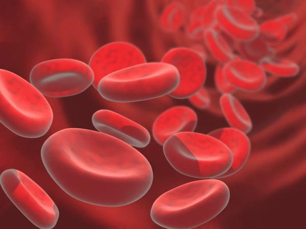
Blood consists of plasma (liquid part of blood) and corpuscles - leukocytes, platelets, erythrocytes. Each type of formed elements has its own functions: leukocytes are responsible for immune protection, platelets - for blood coagulation, erythrocytes provide the transport of oxygen and carbon dioxide.
In a healthy person, the composition of the blood is quite constant, but with illness it changes. Therefore, with the help of a blood test, it can be established that the disease is present. Sometimes a complete blood count can detect the disease on early stagewhen the main symptoms of the disease have not yet been manifested. That is why the UAC is carried out during any preventive examination. In the presence of symptoms, clinical analysis helps to understand the nature of the disease, to determine the intensity of the inflammatory process. Clinical analysis is used to diagnose various inflammatory diseases, allergic conditions, and blood diseases. A repeated general blood test will give the doctor the opportunity to judge the effectiveness of the prescribed treatment, assess the tendency to recovery and, if necessary, adjust the course of treatment.
Indicators of a clinical blood test
A general blood test necessarily contains the following indicators:
If necessary, the doctor may prescribe an extended clinical blood test. In this case, he will specifically indicate which indicators should be additionally included in the analysis.
Decoding of indicators of a general blood test
Hemoglobin
Hemoglobin is a protein that is part of the red blood cell. Hemoglobin binds to oxygen and carbon dioxide molecules, which allows oxygen to be delivered from the lungs to tissues throughout the body, and carbon dioxide back to the lungs. Hemoglobin contains iron in its composition. It is he who gives the red color to erythrocytes (red blood cells), and already those to blood.
The saturation of blood with hemoglobin is an extremely important indicator. If it falls, the tissues of the body receive less oxygen, and oxygen is needed for the life of each cell.
The hemoglobin norm for men is 130-160 g / l, for women - 120-140 g / l. Children are not dependent on gender, but a newly born child has a number of red blood cells (and, accordingly, the level of hemoglobin) significantly exceeds the "adult" norm. And the first 2-3 weeks, this indicator gradually decreases, which must be borne in mind when evaluating the results of a general blood test.
When hemoglobin values \u200b\u200bare below normal, it is diagnosed. Also, a low hemoglobin level may indicate overhydration of the body (increased fluid intake). Hemoglobin above normal, respectively, can be observed with dehydration (blood thickening). Dehydration can be physiological (for example, due to increased physical activity), or maybe pathological. Elevated level hemoglobin is a typical sign of erythremia, a bleeding disorder in which an increased number of red blood cells is produced.
Erythrocytes
Red blood cells are red blood cells. There are significantly more of them than all other shaped elements combined. That is why our blood is red. Erythrocytes contain hemoglobin and thus participate in the process of oxygen metabolism in the body.
The norm for erythrocytes for men is 4-5 * 10 12 per liter of blood, for women - 3.9-4.7 * 10 12 per liter.
Color index
The color index is calculated according to the formula correlating the hemoglobin level and the number of red blood cells. Normally, the color index should be close to one (0.85-1.05). A deviation from the norm is observed with anemia, and with different types of anemia manifests itself in different ways: a color indicator below the norm indicates an iron deficiency (hemoglobin level is reduced to a greater extent than the number of red blood cells); a color indicator above the norm is typical for other types of anemias (the number of red blood cells decreases to a greater extent than the level of hemoglobin).
Reticulocytes
Reticulocytes are young, not yet matured forms of erythrocytes. The process of erythrocyte formation is continuous, therefore reticulocytes are always present in the blood. Norm: 2-10 reticulocytes out of 1000 erythrocytes (2-10 ppm (‰), or 0.2-1%). If reticulocytes over normal, this suggests that the body feels the need to increase the number of red blood cells (for example, due to their rapid destruction or blood loss). A reduced level of reticulocytes is characteristic of anemia, radiation sickness, oncology (if metastases have affected the bone marrow), and some kidney diseases.
Platelets
The main function of platelets is to provide hemostasis, that is, in other words, platelets are responsible for blood clotting. They also participate in the body's immune response to infection. Norm: 180-320 * 10 9 per liter. A lowered platelet count may indicate a severe inflammation or autoimmune disease. An increased level is typical for conditions after significant blood loss (for example, after an operation), and is also observed in cancer or atrophy (decreased function) of the spleen.
Leukocytes
Leukocytes are white blood cells that have a protective function, that is, they represent the immune system. Normally, the total number of leukocytes should be in the range of 4-9 * 10 9 per liter.
An increase in the number of leukocytes indicates the body's immune response and is observed in infectious diseases (primarily caused by bacteria), inflammatory processes, and allergic reactions. A high level of leukocytes can also be a consequence of recent bleeding, stress, tumor processes, and some other pathologies.
A lowered level of white blood cells indicates a depressed state of the immune system. Such results can be observed with a viral infection (,), severe toxicosis, sepsis, diseases of the hematopoietic organs, radiation sickness, autoimmune diseases etc.
It is not only the general assessment of the number of leukocytes that matters. There are five types of leukocytes - neutrophils, eosinophils, basophils, lymphocytes and monocytes; they all have different functions, and therefore it is important to know in what proportion they are present in the blood. The ratio of different types of leukocytes in their total volume is called leukocyte formula.
Neutrophils
An increase in the number of neutrophils in the blood, therefore, indicates the presence of an infection (first of all, one should suspect bacterial infection), the ongoing inflammatory process. It can also be the result of stress, intoxication, cancer.
Eosinophils
Basophils
Norm: 0-1% of the total number of leukocytes.
Lymphocytes
Lymphocytes are the main cells of the immune system. They provide specific immunity, that is, they recognize the penetrated foreign agent and destroy it. With the help of lymphocytes, the body fights viruses. Normally, lymphocytes make up 19-37% of the total number of leukocytes. In children, the proportion of lymphocytes is higher. At the age of 1 month to two years, lymphocytes are the main type of leukocytes, they are the main mass observed. By the age of 4-5, the number of leukocytes becomes comparable to the number of neutrophils. As the child grows up, the decrease continues, but even at the age of 15, children have more lymphocytes than adults.
An increased content of lymphocytes in the blood indicates the penetration of a viral infection; it is also noted with toxoplasmosis, tuberculosis, syphilis.
A lowered lymphocyte count is a sign of a depressed immune system.
Monocytes
Monocytes are in the blood on average for about 30 hours, after which they leave the bloodstream and pass into tissues, where they turn into macrophages. The purpose of macrophages is to finally destroy bacteria and dead body tissues, clearing the site of inflammation for subsequent regeneration (restoration of healthy tissue). The norm for monocytes is 3-11% of the total number of leukocytes.
An increased number of monocytes is characteristic of sluggish and long-term diseases, it is observed in tuberculosis, sarcoidosis, syphilis. It is a specific symptom.
ESR - erythrocyte sedimentation rate
If a test tube with blood is left in upright position, erythrocytes - as a heavier fraction of blood compared to plasma - will begin to settle to the bottom. Ultimately, the contents of the tube will split into two parts: the thick and dark part at the bottom (these will be the red blood cells) and the light part at the top (blood plasma). The erythrocyte sedimentation rate is measured in mm / hour. Norm: 2-10 mm / hour for men and 2-15 mm / hour for women. In children, pregnant women and the elderly, the normal range will be different (in children, it varies greatly with age).
White blood cells are an important part of the body, protecting it from harmful bacteria and substances. They swallow and disarm foreign particles. Consequently, the behavior of these cells can show the presence of an inflammation process, because the composition of the blood shows the state of human health. Therefore, for a diagnosis that provides results about, a special analysis is prescribed, which is used in medicine under the name of the leukocyte blood count. Judging by its results, one can learn about the type of the disease, suggest its course and predict the further outcome. What can show a leukocyte formula?
Indicators
Notifies about changes in some types of leukocytes. Often, such a study is prescribed with general analyzes for routine medical examinations, infectious diseases, and for the control of various diseases.
These are cells of the immune system that are responsible for protecting the human body. Their goal is to form a certain border, beyond which harmful substances, toxins, and foreign bodies should not fall.
There are several types of leukocyte cells that perform a specific task. Basophils, monocytes, neutrophils, eosinophils, lymphocytes constitute the body's defense group. What functions do these cells perform?
This species is responsible for security. They recognize, embrace and destroy viruses or bacteria. They are divided into:
- myelocytes (embryos) and metamyelocytes (derived from myelocytes). Basically, they are not in the blood of a healthy person, but in the case of a serious illness, they appear.
- stab (young) - for infections or diseases that are bacterial in nature, their number increases if segmented are not able to neutralize the infection.
- segmetonuclear (mature) - are in the greatest number, since they constitute the body's defense in a normal state.
Lymphocytes... They create antiviral immunity, as they are able to memorize antigens, and also participate in the synthesis of antibodies.
In their functions, they are similar to neutrophils, but they differ in that they are able not only to capture and destroy harmful bacteria, but also to absorb dying cells. Thus, they cleanse the blood, giving the ability to regenerate tissues.
Basophils... They appear when allergic processes occur that do not allow the spread of harmful microorganisms and toxins throughout the blood.
The leukocyte blood count shows the condition of a sick person, the severity of his disease, the causes and its outcome. In addition to the leukocytogram, there are leukocyte indices, which show the level of protein bodies in the blood.
Such an example is the leukocyte index of intoxication, which determines the bulkiness of the inflammation process. And also other types of indices, for example, immunoreactivity, allergization. They help to assess the level of body resistance, the capabilities of the immune system, the patient's condition.
And finally, a leuco formula is used to determine the balance of these bodies in the blood.
Analysis
Before you start a leukocyte formula, you need to go through not difficult preparation. You just need to give up food for 3-4 hours, and also not be exposed to physical and emotional stress.
The material is blood from a vein. Then it is placed on a special glass plate under a microscope. A laboratory technician fishes out several hundred cells to determine the number and level of white blood cells. The next step is to distribute the blood over the entire glass surface, but not evenly. The heavy bodies are at the edges, and the lungs are in the center. The heavy include: monocytes, basophils and eosinophils, and the lungs are lymphocytes.
When counting white bodies in blood, 2 options are used:
- schilling's method. The counting occurs conditionally in 4 areas of the smear.
- filipchenko's method. The laboratory assistant divides the smear into 3 parts, and determines the quantity in a straight transverse line.
However, there are clinics equipped with new equipment and counting leukocytes is done by a special apparatus - an analyzer. And if the result deviates sharply from the norm, then the person intervenes. It should be noted that there is a quantity error anyway. Factors include errors in blood sampling, preparation of a smear, and others.
Ready in a few days. The attending physician analyzes the obtained values.
A specially trained specialist is responsible for decoding the leukocyte blood count. However, you can also compare the result with the norms. To do this, you need to know what indicators are the maximum permissible for a healthy person in accordance with his age.
There are norms of the leukocyte blood count for adults:

- neutrophils - 55%;
- lymphocytes - 35%;
- monocytes - 5%;
- eosinophils - 2.5%;
- basophils - 0.5%.
Norms of the leukocyte formula by age:
- hemoglobin - a protein found in erythrocytes. It is needed to transport oxygen throughout the body, as well as carbon dioxide. For men: 130 - 160 g / l, for women: 120 - 140 g / l, for children from 0 to 6: 100 - 140 g / l, and up to 12: 120 - 150 g / l.
With a deviation of indicators in the leukocyte formula, for example, in a decreasing direction, possible development or leukemia. If enlarged, it indicates the presence diabetes mellitus, dehydration or diseases of the organs of the hematopoietic system.
- erythrocytes... The norm for men is 4.0-5.0 × 10¹² / l, for women: 3.6 - 4.6 × l, in children from 0 - 6 years old: 5 - 15.5 × l, in children from 0 - 6 years old: 5.0-15.5 × l, up to 12 years - 4.0 - 13.5 × l.
Perhaps with drug allergies, sinusitis, bronchitis, leukemia. If the indicators are less than normal, then this indicates the initial stage of the processes of inflammation, the development of viral or infectious diseases.
- neutrophils. Normal amount the content of segmented neutrophils for adults is from 50 to 70%, for children from 0 to 6: 28 - 55%, up to 12 years old: 43 - 60%. As for stab, in adults 1 - 3%, and in children up to 16 1-5%. A deviation from the norm shows that not everything is in order in the body. So, if the amount is exceeded, then it is mainly during bronchitis, sinusitis, organ inflammation. Reduces this indicator of diseases that are infectious or blood diseases.
In the interpretation of the analysis for the leukocyte formula, there is such a term as a shift in the leukoformula. It characterizes the content of stab and segmented in ba. If the shift is to the right, then there are fewer stab neutrophils than more or less, which is reflected in the state of the human-segment neutrophils. Then the person's condition is associated with impaired liver, kidney function or the presence of megaloblastic anemia. If the shift to the left, then stab increases and metamyelocytes, myelocytes appear. Then such diseases emerge: acidosis or acute infections. Also with physical stress.

- eosinophils. For newborns and infants up to 2 weeks, the norm is 1 - 5%, for infants 1 - 6%, from 1 to 2 years this figure is 1 - 7%, from 2 to 5 it is 1 - 6%, and then the norm remains unchanged 1 - five%. A high level of eosinophils occurs with allergic sensitization, with diseases of an infectious nature, neoplastic or diseases of the hematopoietic system. A decrease occurs in stressful conditions, purulent infections, injuries and burns, intoxication.
- monocytes are responsible for recognition foreign bodies... For newborns, the norm is 3 - 12%, then for a 2 week old baby, the indicator rises from 5 to 15%, in infants 4 - 10%, in children under 2 years old 3 - 10%, and then the indicator does not change. occurs with fungal and viral infections, rheumatic diseases, diseases of the hematopoietic system. It is also possible during the recovery period. A decrease is observed during childbirth, shock conditions, when taking glucocorticoids. Also with aplastic anemia or hairy cell leukemia.
- basophils... The norm is 0 - 0.5% for everyone. An increase in basophils is observed with such diseases: chickenpox, myxedema, chronic myeloid leukemia. For other diseases: Hodgkin's disease, ulcerative colitis, chronic anemia, nephrosis. A decrease in basophils occurs during pregnancy, ovulation, pneumonia, hyperthyroidism, and also with pathologies in the bone marrow.
- lymphocytes... This indicator changes throughout life. For newborns 15 - 35%, for babies up to 2 weeks 22 - 55%, in infants 45 - 70%, in children under 2 years old 37 - 60%, up to 5 years old 33 - 55%, up to 8 years old 30 - 50%, up to 15 years this figure is 30 - 45%, and then unchanged 20 - 40% An increase in lymphocytes indicates ARVI, viral infections, blood diseases, and poisoning. A decrease in lymphocytes is observed in acute infections and diseases, miliary tuberculosis, aplastic anemia, renal failure, HIV - infections.
The leukocyte blood count in children contains some differences depending on age.
For a newborn baby, the ratio of the blood form is stable. However, the number increases by the 6th day to 49 - 60%, and neutrophils decrease to 35 - 48%.
In the first months of life, a leukoformula is formed in a child, which will persist for a whole year. Indicators for infants have some differences in lability, they can be easily violated when the child is indignant or anxious, in case of illness, climatic changes. Up to 6 years, the number of neutrophils and lymphocytes increases. Closer to 15 years, the leukogram becomes similar to that of an adult.
And so, it turned out that the leukocyte blood count in children will naturally change in connection with his age. The number of neutrophils in the blood of a newborn is in the range from 51 to 71%, gradually increases in the first days of life, and then begins to decline sharply. At this time, the baby fluctuates from 15 to 35%, by the end of the second week it reaches 55%. When a baby is 6-7 days old, the curves of lymphocytes and neurophils converge. This intersection is called the first intersection.
As for the basophilic ones, they are almost absent in newborns. The number of monocytes in the blood ranges from 6.5 to 11%, and at the end of the first week from 8.4 to 14.1%. Plasma cells are quite small, from 6.4 to 11.2%. In babies up to a week, there is a clear shift to the left along the Schilling, which is balanced until the end of the week.

For a month of a baby's life, a leukogram is drawn, which will be during the first year. In it, lymphocytes have an advantage, there is always a shift of neutrophils in left side, balanced monocytosis and the presence of plasma cells. Differentiated leukocyte counts in infants fluctuate widely.
When a child is already attending school, their number decreases, and neutrophils increase. Also, the number of monocytes decreases slightly, and plasma cells are no longer present. At the age of 15, the leukogram becomes closer to adults. Accurate estimation of ratios different forms leukocytes in the blood is of great importance in diseases.
How to determine the type of infection
Leukoformula in children and adults gives answers to many questions in diseases of an infectious nature. But how to distinguish between viral and bacterial?
When taking a smear, blood is smeared on the glass. After that, the laboratory doctor takes a microscope, puts it down and looks, observing the behavior of leukocytes. When he saw him, then by appearance determines what kind it is and records the quantity of each type. He does this until he gets 100.
The relationship of different blood cells shows the type of infection. If a large percentage of lymphocytes prevails, then this is a viral infection, if neutrophils, then bacterial.
The main fighter against infections and bacteria is the segmented neutrophil. It is the most popular cell in the blood. In other words, she is mature and ready to deal with all the foreign bodies in her body. If there are many of them, then the body is protected from all bacteria.
However, in order for a segmented neutrophil to become mature, it must undergo a series of transformations. At first he is born in the form of another neutrophil - stab. And when the human body is attacked by any sore, then information is sent to the bone marrow so that the production of young stabs begins. And if there are many of them, then this means that there is an acute bacterial infection.
To educate and secure yourself and, first of all, your baby, in our time, it is possible to undergo many examinations and diagnostics. Especially on the content of leukocytes in the baby's blood. After all, this is very important information about your child's health.
Indications for the analysis
There are a number of necessary cases for taking a blood test:
- must be examined by a doctor once a year
- with complications in diseases
- with fatigue.
ESR analysis allows you to assess a certain rate of sedimentation and separation of blood into plasma and erythrocytes. This method is very effective and reliable, since in the 21st century technology does not stand still and medicine needs high-quality diagnostics of any type of disease or problem of epidemics, etc. The popularity of this analysis has increased, since it is technically simple and affordable, and the results are reliable. But if everything is fine with the indicators, can we assume that the person is not sick? And if the opposite?
Good erythrocyte sedimentation rate results do not mean that the human body is not infected by bacteria or infections. Referring to the data, most patients have ESR less than 20 mm / hour. And in some places, even with increased ESR 100 mm / hour is not possible to learn about the signs of the disease.
 ESR rates according to Westergren
 Therefore, an increase in ESR in the blood in most cases occurs when:
- infections, as processes of an infectious nature increase ESR
- malignant diseases (solitary tumors, etc.)
- rheumatological
- kidney pathology.
For this method, a Panchenkov apparatus is used, which consists of 100 mm pipettes and a tripod. The analysis is performed on the basis of blood from a vein or from a capillary, into which a substance is placed, preventing it from being wrapped. In this case, the smear is placed in a thin test tube and watched for about an hour. The test tube is made of glass or plastic. During this time, there is a division into separate erythrocytes and plasma. ESR is calculated by size from the edge at the top of the plasma to erythrocytes. The normal indicator is a slow erythrocyte sedimentation, followed by the remainder of pure plasma.
There is another “stopped stream” method, in which the sample is stirred to disaggregate the red blood cells. This process must be efficient, otherwise microblocks can change the result. Measurements range from 2 to 120mm / h. The results are highly accurate.
When high level proteins, erythrocytes stick together. Therefore, they go down very quickly, and the ESR in the blood increases its level. As a result, acute or chronic illness can lead to increased ESR. In women, ESR is higher than in men, since there are fewer of them.
ESR rate for adolescents under 15 years old: 2-20mm / hour, from 15 to 50: 2-15 mm / hour, and after 50: 2-20mm / hour. For women allowed values up to 50 range from 2 to 20 mm / hour, and after 50 from 2 to 30 mm / hour.
What is the need
This is necessary to diagnose diseases with an acute or chronic nature, cancer infections. However, this type of analysis is carried out in conjunction with others, since it does not give an exact answer to the type of origin of the disease, its development and outcome.
It is prescribed for monitoring infectious, oncological and autoimmune diseases. And also in combination with a leukocyte blood count or a general blood test.
A clinical blood test can give many answers for complex diagnoses and diseases, as well as describe a person's condition. However, decoding should be done by an experienced specialist who can give an accurate description and correct the treatment process.
Complete blood count (CBC) is a medical study that almost every person has had to deal with. People tend to have a curiosity that they try to satisfy, especially when it comes to their health. In polyclinics, you can often see how a sympathetic therapist explains in detail to the patient all the implications of his analysis.
How to decipher without the help of a specialist a general clinical blood test obtained from a hematological analyzer? It is not enough to read Latin letters and digital symbols - knowledge is needed in order to decipher such information. Fortunately, there is the Internet, and it contains everything you need to decode any information. Online decryption is available on many resources of the world wide web, it can be used by a person who does not have special knowledge.
General (clinical) blood test
What is a complete blood count and why is it called clinical? Complete blood count - diagnostics of the patient's health status using laboratory methods study of blood parameters - white and red cells. Such a blood test is called clinical because this examination is included in the group of general clinical research methods.
In what cases is a clinical analysis prescribed?
The purpose of the general analysis is to provide generalized information about the physiological state of the patient. When a person complains about their health, the doctor examines the patient. The examination procedure is the first stage in the patient's diagnosis. On the basis of the data obtained, the doctor forms a primary clinical picture of the patient's health condition. The second stage is diagnostics based on physiological parameters - blood, feces, urine tests.

The interpretation of the results by the therapist is compared with the conclusions initial examination and as a result, treatment and regimen are prescribed. In cases where the doctor remains in doubt, he can prescribe additional examinations, for example, a biochemical blood test, ultrasound diagnostics, serological analysis, hormone analysis thyroid gland.
With the help of a general analysis, the diagnostician can identify such ailments as:
- leukemia;
- anemia of various types;
- problems with blood viscosity and clotting;
- infectious invasions of various etiologies;
- inflammatory process.

Even a child will be able to describe the procedure for taking blood - a laboratory assistant with a scarifier (a needle for puncturing the skin) pierces a bundle of a finger, brushes the first drop of blood with a cotton swab, then draws blood into test tubes with a glass adapter. In some cases, a laboratory assistant can take material using a vacuum or closed scarifier - such instruments are already found in laboratory practice.
Attention! A detailed clinical analysis suggests actions that require blood of a special quality and in a larger volume, so blood for it can be taken from the cubital (ulnar) vein.
How to properly prepare for a general blood test?
In many medical posts and clinics there are thematic posters and wall newspapers - it is always useful to read them for self-educational purposes. They contain the rules for visiting a doctor on the eve of blood sampling. Usually, people sitting in line for a doctor, trying to somehow occupy themselves, read this information. While the patient has read everything, the queue draws nearer and time passes unnoticed.

Does the age and gender of the patient play a role in decoding a complete blood count?
Decoding a general blood test, in addition to common values, takes into account additional factors - age and gender.
When decoding the values \u200b\u200bin the general blood test, we must pay attention to the person's age - the child's indicators are seriously different from the adult. Children have a different metabolism, different digestion, different immunity, and their blood has a different composition. The situation changes with age. A child ceases to be considered such after hormonal changes in the body: in girls this happens at the age of 11-13; for boys - at 12-14 years old. Moreover, a sufficient period of time is needed to children's organism formed finally. The period of life of children before hormonal changes is called prepubertal in medicine, after - pubertal.

The norms of general analysis for women also have their own characteristics, their difference from men is not very significant, but there are some nuances: a) menstrual cycle; b) gestation (pregnancy).
Attention! Menstruation is a limiting factor for donating blood for a general analysis. The doctor must be warned about monthly cycle and wait for his decision.
Decoding a blood test using a table
The decoding of the clinical blood test is based on normal indicators, thanks to which you can find out about the presence of pathological changes in the patient's body. The norms of the clinical analysis of blood are indicated in the table. There is a separate table for adults (for women and men) and for children.
| Parameters | Index | Units | Range of norms in adults | ||
| In men | Among women | ||||
| Monocytes | * MON * | % | 3,04-11,04 | 3,04-11,04 | |
| Lymphocytes | * LYM * | % | 19,43-37,43 | 19,43-37,43 | |
| Leukocytes | * WBC * | 10 9 cells / l | 4,02-9,01 | 4,02-9,01 | |
| Basophils | * BAS * | % | 0,1-1,0 | 0,1-1,0 | |
| Neutrophils | stab | % | 1,01-6,10 | 1,01-6,10 | |
| segmented | % | 46,80-66,04 | 46,80-66,04 | ||
| * RBС * | х10 12 cells / l | 4,44-5,01 | 3,81-4,51 | ||
| Eosinophils | * EOS * | % | 0,51-5,03 | 0,51-5,03 | |
| Color index | * CPU * | — | 0,81-1,03 | 0,81-1,03 | |
| * PLT * | 10 9 cells / l | 180,0-320,0 | 180,0-320,0 | ||
| Thrombokrit | * PCT * | % | 0,12-0,41 | 0,11-0,42 | |
| ESR | * ESR * | mm / hour | 1,51-10,51 | 2,11-15,11 | |
| Hemoglobin | * Hb * | g / l | 127,0-162,0 | 119,0-136,0 | |
| Hematocrit | * HCT * | % | 128,03-160,03 | 117,0-137,0 | |
Attention! The information in the tables is published for informational and self-educational purposes only. It is approximate and cannot be a reason to start self-medication. If a person is sick, he should see a doctor!
| Parameters | Units | Normal values \u200b\u200bfor children | ||||
| first days of life | up to 1 year | from 1 to 6 years old | from 6 to 12 years old | from 12 to 16 years old | ||
| Reticulocytes | ppm | 3,1-15 | 3,1-12 | 2,1-12 | 2,1-11 | 2,1-11 |
| ESR | mm / hour | 0,11-2,01 | 2,01-12,0 | 2,01-10,0 | 2,01-10,0 | 2,01-10,0 |
| Thrombokrit | % | 0,16-0,36 | 0,16-0,36 | 0,16-0,36 | 0,16-0,36 | 0,16-0,36 |
| 10 9 cells / l | 181,50-400 | 181,50-400 | 181,50-400 | 157,10-380 | 157,10-387,50 | |
| % | 0,83-1,13 | 0,73-0,93 | 0,83-1,10 | 0,83-1,10 | 0,83-1,10 | |
| Eosinophils | % | 2,10-7,14 | 1,10-6,14 | 1,10-6,14 | 1,10-6,14 | 1,14-5,10 |
| х10 12 cells / l | 4,40-6,60 | 3,60-4,92 | 3,50-4,52 | 3,50-4,72 | 3,60-5,20 | |
| Segmented neutrophils | % | 30,10-50,10 | 15,10-45,10 | 25,10-60,14 | 35,10-65,21 | 40,10-65,21 |
| Stab neutrophils | % | 0,52-4,11 | 1,10-5,01 | 1,11-5,0 | 1,11-5,0 | 1,11-5,0 |
| Basophils | % | 0-1 | 0-1 | 0-1 | 0-1 | 0-1 |
| Hemoglobin | g / l | 137-220 | 98-137 | 108-143 | 114-148 | 114-150 |
| Leukocytes | 10 9 cells / l | 7,22-18,50 | 6,14-12,04 | 5,10-12,0 | 4,41-10,0 | 4,33-9,51 |
| Lymphocytes | % | 22,12-55,12 | 38,12-72,12 | 26,12-60,12 | 24,12-54,12 | 25,12-50,12 |
| Monocytes | % | 2,0-12 | 2,0-12 | 2,0-10 | 2,0-10 | 2,0-10 |
Attention! In the tables, the most common units of measurement for the results of a complete blood count were given. Some research medical centers these values \u200b\u200bcan vary, which are indicated with respect to the qualitative and quantitative component of the study. Because of this, deciphering the results must be done carefully.
General clinical blood test parameters
The indicators of a general blood test can be conditionally divided into three groups: leukocytes, erythrocytes and platelets. Each of these groups has its own subgroups: in the first - granulocytic (basophils, eosinophils, neutrophils) and agranulocytic (lymphocytes and monocytes); in the second - erythrocytes plus ESR, hemoglobin plus hematocrit and color index; in the third - platelets plus thrombocyte.

Leukocytes
| Parameter | Description | Increased blood level | Low blood level | Notes |
| Leukocytes | The standard blood count for leukocytes is 4-9 per 10 9 cells / liter. Leukocytes are the general name for all white blood cells. The parameter is needed to determine the number of white cells in human blood. An increased level of leukocytes is called leukocytosis, a decreased level is called leukopenia. | The overwhelming majority of infectious diseases, various internal inflammations, after eating, after vaccinations, during menstruation, the development of oncological pathology (with some types of leukemia, the level of leukocytes in the blood decreases), a good diet. | An insignificant part of infectious diseases (immunodeficiency syndrome, consumption), radiation injuries of all types (solar radiation, radiation therapy, radiation exposure), leukemia (some forms of reticulosis), poor diet. | The parameter gives the most general information about the nature of the disease. According to the indicator, it is impossible to accurately establish the cause of the disease, only its presence. All pathologies indicated in the sections of high and low levels apply to all types of leukocytes. |
| Granulocytes | ||||
| Eosinophils | Microphages. They carry granules with Ig E. They have the ability to attack antigens with histamine, so eosinophils are one of the causes of allergies, but at the same time, these cells can absorb histamine and prevent allergies. | Autoimmune reactions, infections, after blood transfusion, after vaccinations, helminthiasis, leukemia and other oncological diseases. | Heavy metal poisoning
reticulosis, radiation injuries of all types, sepsis, chemotherapy, rheumatism. |
|
| Basophils | The largest of the granulocytes are white blood cells. Their amount in the blood of a healthy person is negligible. Contains histamine, serotonin and other powerful biological irritants, allergic and allergic reactions. Microphages. | Autoimmune diseases of varying intensity, rheumatoid factor, allergic reactions, thyroid and parathyroid glands, nephritis and other inflammatory lesions of the kidneys, gestation with Rh-conflict, rehabilitation after surgical removal of the spleen, after blood transfusion, after vaccinations, during nematodosis (enterobiasis, ascariasis and others), leukemia, the consequence of taking corticosteroids, stomach and duodenal ulcers ... | Not | Since a healthy person should not normally have basophils in the blood, pathologies of a reduced level are not indicated. |
| Neutrophils | They are divided into 2 types - stab and segmented. Microphages. The most common of all leukocytes - the number of the total mass of leukocytes is 70%. | Bacterial infections, leukemia, uremia, diabetes (sugar),taking immunostimulants | Viral infections, reticulosis, with hyperethyroidism, with radiation injuries of all types, after chemotherapy. | |
| Agranulocytes | ||||
| Monocytes | The largest type of leukocytes. Macrophages. | Allergies, infections, leukemia, phosphorus isoform poisoning. | Reticulosis and hairy cell leukemia, sepsis. | |
| Lymphocytes | Fighters of the body number 1. They resist any biological and non-biological threats. They are divided into three main types - T-lymphocytes (75% of all lymphocytes), B-lymphocytes (15%) and null cells (10%). | Infectious invasions of various origins, leukemia,poisoning with heavy metals (lead, mercury, bismuth, arsenic), preception of immunostimulants. | Consumption, immunodeficiency syndrome,reticulosis, radiation injuries of all types, chemotherapy, rheumatism. | |
Erythrocytes, hemoglobin, hematocrit, ESR, color index
Erythrocytes are red blood cells. Visually, these are scarlet plates, concave in the middle. The shape of red blood cells that we have described is the shape of normal red blood cells; there are forms that indicate pathological abnormalities in the structure of red blood cells as a result of severe hereditary diseases, infection (sickle erythrocytes - a symptom of the development of malaria), abnormalities in the work of metabolism. The red color of erythrocytes is given by the pigment protein hemoglobin, its main property is the retention of iron atoms in its structure. Thanks to iron, hemoglobin is able to bind oxygen and oxygen oxide - this ability allows metabolic processes in cells. Oxygen is an important participant in many biochemical processes in the body.
 General analysis, studying the state of erythrocytes, is interested, first of all, how much hemoglobin is in the erythrocyte. For this, ESR and color index methods have been developed. ESR - which means "erythrocyte sedimentation rate". Hemoglobin is a heavy protein, and if blood is drawn into a test tube, then, after an hour, erythrocytes will go down in relation to the extracellular fluid. From the rate of subsidence and the depth of subsidence of red cells, it can be concluded how much hemoglobin is contained in erythrocytes and what quality it is - normal or defective. There are no clear standards in this procedure - further diagnosis will depend on the interpretation of other clinical data.
Attention! The mass fraction of red blood cells relative to a unit volume of blood is called hematocrit.
The color indicator also examines the hemoglobin content in erythrocytes. A laboratory assistant, examining erythrocytes under a microscope, looks at the center of the red cell (hemoglobin is concentrated there): if the erythrocyte has a transparent center, this will indicate the absence of hemoglobin in the cell or dysfunction of the peptide chain (hypochromia); if the center is orange, hemoglobin is normal (normochromia); if the center of the cell merges in color with the body of the erythrocyte - excess hemoglobin (hyperchromia).

Platelets, thrombocyte

Platelets are cells that play an important role in the blood clotting process. Platelets do not have a nucleus. Structurally, platelets are a piece of the cytoplasm of megakaryocytes, so their study gives a lot of information about the state of the bone marrow. The number of platelets in the blood, their qualitative composition is an important clinical marker of the bone marrow function.
The norms of a general blood test for platelets are 180-320 in 10 9 cells per liter. The total number of platelets, like erythrocytes, is measured in absolute values \u200b\u200brelative to a unit volume of blood. This parameter is called "thrombocrit".
From the article, the reader learns what a general blood test shows, in which cases it is prescribed, which indicators include a general analysis. How to prepare for the test delivery procedure, and what factors can affect the results. You will learn the normal values, how they change under various conditions and diseases of the body.
A blood test is an important stage in examination and diagnosis. The hematopoietic organs are susceptible to physiological and pathological influences. They change the blood picture.
As a consequence, the general analysis (GAC) is the most popular analysis methodwhich helps the doctor judge general condition organism. For a detailed examination, in addition to the CBC, a biochemical analysis and a general urine test (OAM) are prescribed. What it shows general analysis of urine, a separate article has already been written. If you are interested, you can read it.
What does the general blood test show, detailed, the main indicators
Let's find out what a general blood test shows, why it is taken. General hematological blood test is an important diagnostic criterion that reflects the response of the hematopoietic system to the action of physiological and pathological factors.
UAC is of great importance in establishing a diagnosis, especially in diseases of the hematopoietic organs. UAC covers the study of such indicators:
- hemoglobin (Hb) level
- erythrocytes
- leukocytes
- platelets
- color indicator
- calculation of leukoformula
- erythrocyte sedimentation rate
If necessary, the clotting time, the duration of bleeding are examined. In many laboratories, the analysis is carried out on hematology automatic analyzers. They define up to 36 parameters at once.

Hemoglobin, function and clinical significance
Hb, a blood pigment, is a core component of an erythrocyte. Its role is to transport O 2 from the lungs to organs, tissues, and the removal of carbon dioxide.
The hemoglobin level plays a major role in the diagnosis of anemias of various etiologies. At the same time, its performance decreases.
An increase in Hb concentration occurs with erythremia, symptomatic erythrocytosis, congenital heart disease, cardiopulmonary failure. An increase in Hb is combined with an increase in the number of erythrocytes.
When acute blood loss there is a significant decrease in Hb to 50 g / l... The minimum content of pigment in the blood compatible with life is 10 g / l.
If you have problems with back pain, I propose to find out what it is, which is also very useful, which is also disclosed in the article - follow the link.
Erythrocytes, physiological role in the body
Erythrocytes occupy the main share in the mass of blood corpuscles, it contains hemoglobin. The main function is the transfer of O 2 with the assistance of Hb. In addition, erythrocytes are involved:
- in the absorption of lipids, amino acids, toxins
- in enzymatic processes
- when regulating acid-base balance of the body
- in regulation of ionic equilibrium of plasma
A decrease in the number of red blood cells is one of the signs of anemia. In addition to anemia, red blood cells decrease with an increase in blood volume in the bloodstream, for example, during pregnancy.
An increase in the number of red blood cells (erythrocytosis) is characteristic of erythremia. KLA in newborn babies will show erythrocytosis within the first 3 days of life. In adults, erythrocytosis is observed during starvation, profuse sweating, and ascents.
Leukocytes and their physiological role in the body
The number of leukocytes (L) in the bloodstream is an important diagnostic criterion. They perform important functions - protective, trophic and others. An increase in the number of leukocytes more than 10 × 10 9 / l (G / l) is called leukocytosis.
Most often, leukocytosis occurs as a result of acute infections caused by cocci. Therefore, the KLA will definitely show inflammation, pneumonia, and blood cancer. Leukocytosis is typical for:
- leukemia of various courses, malignant tumors
- inflammatory, purulent, acute infectious processes
- uremia
- myocardial infarction
- toxic poisoning, severe blood loss, shock conditions, extensive burns
UAC at acute appendicitis will show an increase in the amount of L. Leukocytosis is characteristic of tubal pregnancy, ruptured spleen, acute gout.
A decrease in the number of leukocytes below 3.5 g / l is called leukopenia... The tendency to leukopenia occurs among healthy population and is often hereditary, but may be influenced by external factors environment (sun radiation).
Sometimes it occurs during fasting, with a decrease in tone, in a dream. Leukopenia is typical for:
- infections caused by viruses and bacteria - typhoid fever, endocarditis, salmonellosis, measles, influenza, rubella
- lupus erythematosus
- hemoblastosis
- and children (read more by clicking on the link)
The appearance of leukopenia is associated with inhibition of cell maturation and the release of L from the hematopoietic organs and their redistribution in the vascular bed.
The diagnostic value of calculating the leukoformula is enormous in many pathological conditions. It can be used to judge the severity of the situation, the effectiveness of the prescribed therapy.

Leukocytes include cells of the lymphocytic, monocytic, granulocytic series. To find out their number use counting leukocyte formula -% content different types leukocytes:
- stab and segmented neutrophils
- eosinophils
- monocytes
- basophils
- lymphocytes
Neutrophils carry out bacteri-candida and virucidal function. They are capable of phagocytosis in capillaries and are involved in all stages of inflammation. Therefore, an increase in the number of neutrophils will show inflammation in the body. Neutrophilia (above 8 × 10 9 / l) is present in any suppurative process, sepsis.
Eosinophilshave a detoxifying effect. They are found in large quantities in tissue fluid, intestinal mucosa, skin.
Eosinophilia accompanies disease connective tissue - polyarteritis, rheumatoid arthritis, tumors, especially with metastases and necrosis.
Eosinopenia (decrease) is typical for an infectious-toxic process, in postoperative period... And indicates the severity of the condition.
Basophilshave anticoagulant properties. Involved in inflammatory and allergic processes. Basophilia occurs with an allergic reaction to food intake, drugs, foreign protein. In oncology - chronic myeloid leukemia, myelofibrosis, erythremia, lymphogranulomatosis.
Typical for ulcerative colitis, estrogen treatment. Basophilia is likely during ovulation and pregnancy, with lung cancer, anemia of unknown origin, and iron deficiency.
Monocyteshave the ability to phagocytosis. They actively phagocytose (absorb) the remains of cells, small foreign bodies, plasmodium malaria, mycobacterium tuberculosis.
With tuberculosis, monocytosis is observed in the blood - an increase in the number of monocytes. Monocytopenia is observed with hypoplasia of hematopoiesis.
Lymphocytesimportant for immunity. In addition, lymphocytes take part in the fight against infection, as well as carry out a trophic function at the sites of inflammation and wounds. Lymphocytosis is possible with infectious mononucleosis, tuberculosis, syphilis.

Platelets - physiological role, clinical significance
Corpuscular element of blood, is involved in hemostasis processes. Thrombocytosis (increase in the number of tr) can be observed under physiological conditions after exercise, due to arousal nervous system... Thrombocytosis occurs when:
- injuries with muscle damage
- burns, asphyxia, after blood loss and removal of the spleen
- leukemia - erythremia, myeloid leukemia
Thrombocytopenia (decrease in the tr number) in physiological conditions occurs during menstrual blood loss in women, after histamine. In pathological conditions, thrombocytopenia occurs when:
In this case, the autoimmune factor is of great importance - the formation of antibodies to their platelets.
Erythrocyte sedimentation rate
An increase in ESR can occur under physiological conditions - during pregnancy, during fasting when eating dry food, after vaccination, while taking certain medications.
Change in ESR in pathology has diagnostic and prognostic meaning... And it serves as an indicator of the effectiveness of the healing process. ESR increases with:
- infections and inflammations
- purulent processes
- rheumatism
- kidney disease, liver disease ( including at)
- myocardial infarction, malignant tumors, anemias
Reduced eSR indicators are found in processes accompanied by blood thickening. Sometimes observed with neuroses, epilepsy, anaphylactic shock, with erythremia.

Total red blood cell volume (hematocrit)
Hematocrit (Ht) is the ratio of plasma to corpuscles. An increase in Ht occurs in heart defects and is accompanied by cyanosis, with erythrocytosis.
A decrease in hematocrit is typical for various anemias in the second half of pregnancy.
Color index
The color or color indicator is the relative amount of Hb in the erythrocyte. A decrease in this value occurs with iron deficiency.
An increase in the color index is observed with anemia, deficiency of Vit B 12 (cyanocobolamine), folic acid. It accompanies cirrhosis of the liver, thyroid disease, occurs during therapy with cytostatics, taking contraceptives, and using anticonvulsants.
Normal blood laboratory test values
An important stage in assessing the result of the UAC is to establish the difference between pathology and the norm. For this, a definition of normal indicators should be given - these are indicators found in healthy people... They may differ depending on gender.
| Index | Normal values | |
| men | women | |
| Hemoglobin, Hb | 125 - 170 g / l | 105 – 155 g / l |
| Erythrocytes, Er | 3.8 - 5.5 T / L | 3.5 - 4.9 T / L |
| Leukocytes, L | 3.8 - 9.5 G / L | |
| Hematocrit | 40 – 50 % | 38 – 47 % |
| ESR | 1 - 10 mm / h | 2 - 12 mm / h |
| Platelets, tr | 150 - 380 × 10 9 / l | |
Segmented neutrophils Stab neutrophils Lymphocytes Monocytes Eosinophils Basophils |
||
When evaluating test results, it should be remembered that deviations outside the norm do not necessarily indicate the presence of a disease.
When interpreting the results, it is necessary to find out if the deviations are of a physiological nature. We should not forget about the variability of the norm associated with personality characteristics.
When interpreting the results, it is required to take into account many factors: age, gender, concomitant diseases, intake drugs, living conditions and much more. Therefore, a doctor should do this.
Place of blood sampling for research: from a vein or from a finger
The results of laboratory studies are significantly influenced by the location and technique of taking biological material. IN medical practice more often use blood from the capillaries. Usually it is taken from the flesh of the ring fingers, in difficult cases - from the earlobe.

The puncture is done on the side, where the capillary network is thicker. The blood should flow by gravity so that there is no mixing of tissue fluid, which will lead to a distortion of the result. For research, capillary blood should be taken:
- with extensive burns of the body, especially hands
- if small or inaccessible veins, obesity
- in patients prone to thrombosis
- in newborns
Currently, blood from the venous bed is revered the best material for general clinical analysis. This is due to the use of hematological analyzers. With their help, in our time, the UAC is carried out. They are designed and standardized for the treatment of venous blood.
When taking blood from a vein, it is also required to follow some rules. The best place to draw blood is in the ulnar vein. A tourniquet should not be applied for more than 2 minutes, this will lead to an increase in cellular elements in the bloodstream.
When evaluating test results, you need to take into account a number of factors that affect them. Let's name the most essential:
- reception and composition of food, diet
- physical stress has a transient and lasting effect on results
- nervous stress increases leukocytosis
- medications
- body position during the taking procedure
- location and technique of blood collection
- time and conditions of delivery of biomaterial to the laboratory
Other factors influencing the results include the patient's age, gender, and ambient temperature. The harmful tendencies - smoking and alcohol - have a great influence. They lead to an increase in the concentration of Hb and the number of red blood cells. On the contrary, the leukocyte count decreases.

Basic rules for preparing for the delivery of the UAC
- cancel as agreed with the doctor medicines the day before the test
- do not donate blood after physiotherapy, X-ray examination
- do not donate blood immediately after mental and physical exertion
- refrain from smoking 1 hour before the procedure
- give up fatty and spicy foods, alcohol 48 hours before the procedure
- go to bed at the usual time, get up no later than one hour before blood sampling
Repeated examinations should be carried out at the same hours, since the morphological composition of the blood is prone to daily fluctuations.
I suggest watching a video of how a complete blood count is done:
Do not neglect the rules of preparation for the research procedure, and false results you are not afraid!
So, now the reader knows what the general blood test shows, the purpose of its appointment, what indicators the general analysis includes. How to prepare for the test delivery procedure, and what factors affect the results. We learned the normal values, how they change under various conditions and diseases of the body.
Still have questions? Ask in the comments.
A general (or clinical) blood test is performed to identify changes in the quantitative and qualitative indicators of its composition. Such a laboratory study of blood components can be carried out for prophylactic purposes to exclude latent indolent diseases, to confirm or refute a preliminary diagnosis, to track the dynamics of the development of an already confirmed disease. Decoding of the results of clinical analysis in adults has the form of a table containing the names of indicators, units of measurement, norms and actually detected deviations in the composition of the blood.
Human blood consists of plasma (liquid part) and shaped elements (cells): leukocytes, platelets, erythrocytes. Their amount in the blood directly depends on the age and sex of the person, as well as his physical condition. Each type of shaped elements has its own functions:
- leukocytes are responsible for immune protection,
- platelets - for blood clotting,
- erythrocytes provide oxygen and carbon dioxide transport.
Most of the processes affecting the state of various tissues and organs, one way or another, are reflected in the composition of the blood. This is evidenced by a change in a number of indicators determined during clinical analysis.
A clinical blood test includes counting all types of cells (erythrocytes, leukocytes, platelets), determining their parameters (size and shape of cells), leukocyte formula, measuring hemoglobin levels, determining the ratio of cell mass to plasma (hematocrit). Also, in the course of the study, ESR (erythrocyte sedimentation rate) is determined, which is a clear indicator of either autoimmune diseases.
IN laboratory diagnostics a general clinical blood test is one of the first places among other laboratory diagnostic procedures.
Indications for analysis
Changes in blood composition can be of diagnostic value in a number of human diseases.
A detailed clinical blood test is a standard study that is used in laboratory diagnostics to identify:
The main function of neutrophils is the formation of immunity. They have detoxification, antioxidant and bactericidal activity and are involved in the formation of an immune response in infectious diseases provoked by pathogenic or opportunistic bacteria.
In the interpretation of the analysis, neutrophils are designated as NEUT%, are determined as a percentage of the total volume of leukocytes. Normally, neutrophils in the blood of adults should contain 45-70%.

An increase in the number of neutrophils - neutrophilia - is evidence of acute bacterial or fungal infections, bleeding, diseases accompanied by tissue necrosis, and malignant neoplasms.
Neutropenia is a low level of neutrophils, indicating a depression of the immune system. Develops as a result of transferred viral infections, other inflammatory diseases in severe form, with anemia or as side effect some medications.Lymphocytes
Lymphocytes are the main cells of the immune system, providing the production of antibodies necessary for the formation of an immune response when in contact with pathogens.
The norm of lymphocytes in women and men does not differ, does not depend on age and is 19-37%.
An excess of the indicator - lymphocytosis - is typical for most viral infections (influenza, SARS, herpes, hepatitis, etc.), exacerbation of allergic diseases.
A low number of lymphocytes is observed with immunodeficiency while taking immunosuppressants, glucorticoids, as well as with some types of anemia and leukemia.
Eosinophils
A variety of leukocytes that have phagocytic properties and are involved in the formation allergic reactions when the body comes into contact with external pathogens.
Normal indicators of eosinophils in the blood in adults do not depend on sex and age and are in the range of 0-5%.
A decrease in eosinophils indicates acute infectious diseases, inflammatory processes in the organs abdominal cavity, blood poisoning. A significant deviation from the norm is observed within 16 hours after the onset of myocardial infarction, surgical intervention, burn or traumatic shock.
Monocytes
Monocytes belong to agranulocytes from the system of phagocytic mononuclear cells (macrophages) - long-lived cells, the properties and functions of which have much in common with neutrophils. They remove old, destroyed and dying cells, antigen complexes and altered native protein molecules from the body.
A decrease in the level of monocytes - a state of monocytopenia - is usually associated with inhibition of the hematopoiesis process against the background of a deficiency of iron, B vitamins, folic acid, as a result of chemoradiation and hormonal therapy.
Basophils
Basophilic leukocytes are the first to react to the appearance of allergens, infections or other damaging factors in the body. They activate the mechanisms of an inflammatory-allergic nature, attracting other types of leukocytes, increasing the reactivity of the vascular wall, smooth muscles, changing the function of the cardiovascular and respiratory system, kidneys.
Normally, the relative number of basophils in the blood of an adult does not exceed 1% of the total number of leukocytes.
An increase in the indicator indicates the presence of food, seasonal or drug allergies, hypothyroidism, chronic inflammatory or autoimmune diseases.
A decrease in basophils can be the result of chronic stress, prolonged use of antibiotics, cytostatics, chemotherapy or radiation therapy.
Platelets (PLT)
Thrombocytes are small, flattened, colorless blood cells that form in the red bone marrow. Platelets are involved in blood clotting. They protect the walls of blood vessels from mechanical damage and prevent significant blood loss.
At rest, platelets are the smallest blood cells. However, if the vessel is damaged under the influence of biologically active substances they are capable of a rapid transition to a new state.
When activated, platelets change their shape - a lot of processes are formed on the surface of the cells, exceeding the size of the platelets themselves. This allows the cells to stick together and attach to the vessel wall, blocking the site of damage to the vascular wall. Thus, if necessary, platelets "close" wounds and stop bleeding.
Platelet counts are recommended for people who suffer from unexplained bruising, bleeding gums, excess blood during menstruation, nosebleeds, and those who have bleeding from a small wound for a long time.
The platelet count is determined in the number of billions of cells per liter of blood (* 10 9 l).
Platelet count in men by age
Platelet count in women by age
A decrease in the number of platelets in the blood can lead to bleeding. An increase in their number leads to the formation of blood clots (thrombosis), which can block blood vessels and lead to pathological conditions such as stroke, myocardial infarction, pulmonary embolism or blockage. blood vessels in other organs of the body.
The average platelet volume is lowered - what does it mean
What this means if the average platelet volume is low (designated as MPV), it is necessary to know people at risk of developing diseases of the thyroid gland, heart, blood vessels. Such clinical picture can be observed with oncological diseases, iron deficiency anemia, pathologies of the hematopoietic system.
Some kidney diseases (for example, glomerulonephritis, an autoimmune, infectious, or allergic inflammation of the renal glomeruli) are also associated with a decrease in the mean platelet count. A physiological decrease in MPV is observed in pregnant women and lactating mothers.
If the average platelet volume is significantly below the physiological norm for several studies in a row, the cause may be oncological diseases, therefore, such patients are required to consult an oncologist.
The average platelet volume is increased - what does it mean
When a patient hears that he has an increased average platelet count, do not panic: first you need to figure out what this means and how dangerous it can be to health.
- The following pathologies can lead to an increase in MPV in patients of any age:
- various forms of anemia;
- helminthic invasion;
- infectious and inflammatory diseases;
- cancerous tumors of the gastrointestinal tract.
In some diseases of the hematopoietic system in the blood, there may also be a significant increase in the average volume of platelets.
General clinical blood test is the most important element primary diagnosisrequired for early detection of existing violations and initial stages inflammatory processes.
It is necessary to donate blood at least once a year. People who are at risk for any pathology or who have chronic diseases should have their blood counts checked 2 to 4 times a year.